Case Reports
. 2013 Aug 15;22(16):3250-8.
doi: 10.1093/hmg/ddt179. Epub 2013 Apr 15.

ALDH1A3 loss of function causes bilateral anophthalmia/microphthalmia and hypoplasia of the optic nerve and optic chiasm

Affiliations
Case Reports

ALDH1A3 loss of function causes bilateral anophthalmia/microphthalmia and hypoplasia of the optic nerve and optic chiasm

Mani Yahyavi et al. Hum Mol Genet. .

Abstract

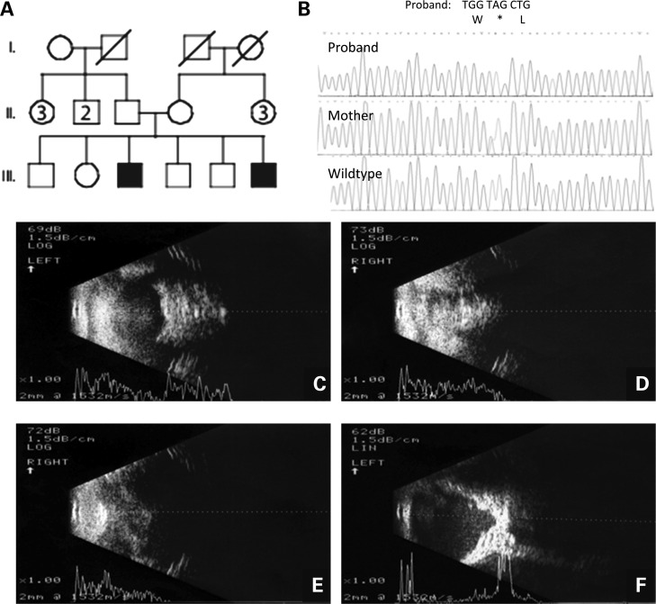
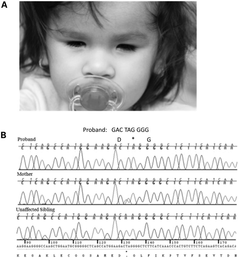
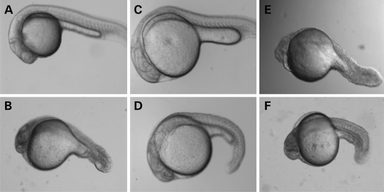
The major active retinoid, all-trans retinoic acid, has long been recognized as critical for the development of several organs, including the eye. Mutations in STRA6, the gene encoding the cellular receptor for vitamin A, in patients with Matthew-Wood syndrome and anophthalmia/microphthalmia (A/M), have previously demonstrated the importance of retinol metabolism in human eye disease. We used homozygosity mapping combined with next-generation sequencing to interrogate patients with anophthalmia and microphthalmia for new causative genes. We used whole-exome and whole-genome sequencing to study a family with two affected brothers with bilateral A/M and a simplex case with bilateral anophthalmia and hypoplasia of the optic nerve and optic chiasm. Analysis of novel sequence variants revealed homozygosity for two nonsense mutations in ALDH1A3, c.568A>G, predicting p.Lys190*, in the familial cases, and c.1165A>T, predicting p.Lys389*, in the simplex case. Both mutations predict nonsense-mediated decay and complete loss of function. We performed antisense morpholino (MO) studies in Danio rerio to characterize the developmental effects of loss of Aldh1a3 function. MO-injected larvae showed a significant reduction in eye size, and aberrant axonal projections to the tectum were noted. We conclude that ALDH1A3 loss of function causes anophthalmia and aberrant eye development in humans and in animal model systems.


Figures

Figure 1.
Figure 1.
Pedigree, chromatograms and transpalpebral ultrasonography from family with ALDH1A3 loss-of-function mutation p.Lys190*. (A) Pedigree of family with two sons affected by A/M. The oldest, III-3, has bilateral anophthalmia and the youngest, III-6, has anophthalmia of the right eye and severe microphthalmia of the left eye. (B) The chromatogram of the oldest son showing c.568A>T, predicting p.Lys190* and premature protein truncation of ALDH1A3. (C) Transpalpebral ultrasonography of the right eye of III-3 showed total anophthalmia without cyst. (D) Transpalpebral ultrasonography of the left eye of III-3 showed total anophthalmia without cyst. (E) Transpalpebral ultrasonography of the right eye of III-6 showed total absence of the eye. (F) Transpalpebral ultrasonography of the left eye of III-6 showed a very small globe with posterior coloboma, detached retina and the presence of the optic nerve in the left orbit.
Figure 2.
Figure 2.
Chromatograms and the phenotype from a simplex case with ALDH1A3 loss-of-function mutation p.Lys389*. (A) Facial view of the patient (simplex case) at 4 years and 6 months of age, showing bilateral anophthalmia. (B) The chromatogram of the patient (simplex case) showing c.1165A>T, predicting p.Lys389* and premature protein truncation of ALDH1A3. Both parents are heterozygous for the mutation, and an unaffected sibling was wild-type.
Figure 3.
Figure 3.
Phenotype in D. rerio larvae injected with antisense MO for Aldh1a3 with rescue experiments. (A) Uninjected D. rerio larva at 24 h.p.f. (B) Representative D. rerio larva injected with translational antisense MO (ATG-MO) for Aldh1a3 at 24 h.p.f. The eye is reduced in size, edema is present and the tail is shortened. (C) Representative D. rerio larva injected with control at 24 h.p.f. (D) Representative D. rerio larva injected with translational antisense morpholino (ATG-MO) for Aldh1a3 together with wild-type human mRNA for ALDH1A3 at 24 h.p.f. Rescue of the morphant phenotype can be observed. (E) Representative D. rerio larva injected with translational antisense morpholino (ATG-MO) for Aldh1a3 and mutant human mRNA for ALDH1A3/p.Lys190* at 24 h.p.f. Rescue of the morphant phenotype is not observed. (F) Representative D. rerio larva injected with translational antisense morpholino for Aldh1a3 and mutant human mRNA for ALDH1A3/p.Lys190* at 24 h.p.f. Rescue of the morphant phenotype is not observed.
Figure 4.
Figure 4.
Aldh1a3 morphant D. rerio larvae show reduced innervation of the tectum. (A and B) Confocal images of Pou4f3:mGFP-labeled retinotectal projections at 5 d.p.f. tecta in live larvae. The wild-type tectum (A) is filled with retinal axons, and the optic tract has branched into stereotyped fascicles. The MO-treated tectum (B) appears less innervated. Figures are shown with the anterior aspect of the larvae superiorly. Dotted lines outline the tectal neuropil.

References

    1. Kawaguchi R., Yu J., Honda J., Hu J., Whitelegge J., Ping P., Wiita P., Bok D., Sun H. A membrane receptor for retinol binding protein mediates cellular uptake of vitamin A. Science. 2007;315:820–825. - PubMed
    1. Duester G. Keeping an eye on retinoic acid signaling during eye development. Chem. Biol. Interact. 2009;178:178–181. - PMC - PubMed
    1. Rhinn M., Dollé P. Retinoic acid signalling during development. Development. 2012;139:843–845. - PubMed
    1. Kumar S., Duester G. SnapShot: retinoic acid signaling. Cell. 2011;147:1422. - PMC - PubMed
    1. Ghyselinck N.B., Dupé V., Dierich A., Messaddeq N., Garnier J.M., Rochette-Egly C., Chambon P., Mark M. Role of the retinoic acid receptor beta (RARbeta) during mouse development. Int. J. Dev. Biol. 1997;41:425–447. - PubMed

Publication types

MeSH terms

Substances