. 2013 Apr 30;110(18):7512-7.
doi: 10.1073/pnas.1301027110. Epub 2013 Apr 16.

Tertiary model of a plant cellulose synthase

Affiliations

Tertiary model of a plant cellulose synthase

Latsavongsakda Sethaphong et al. Proc Natl Acad Sci U S A. .

Abstract

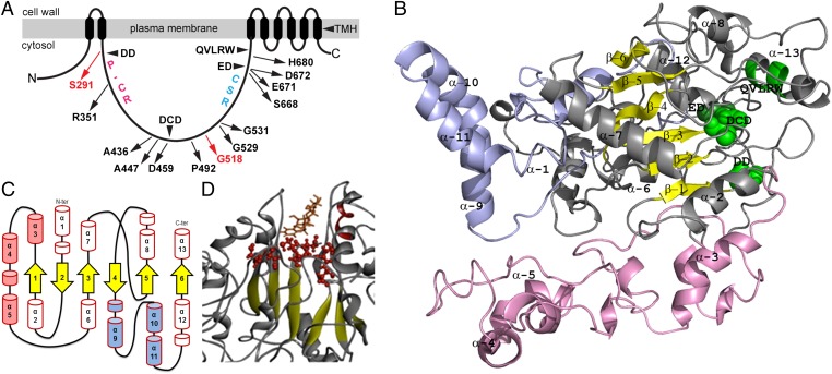
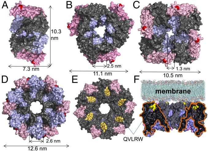
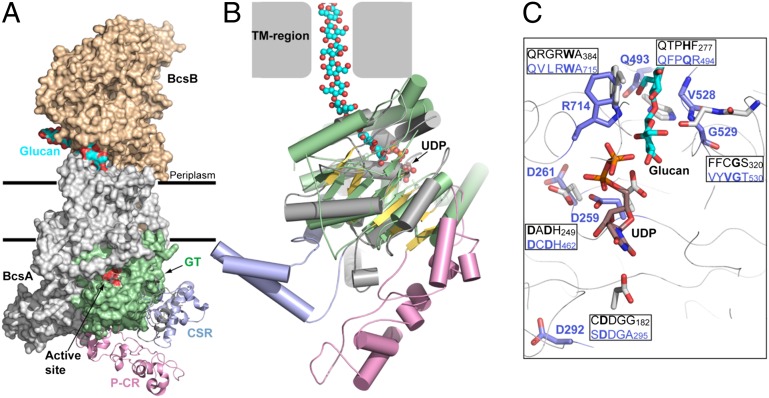
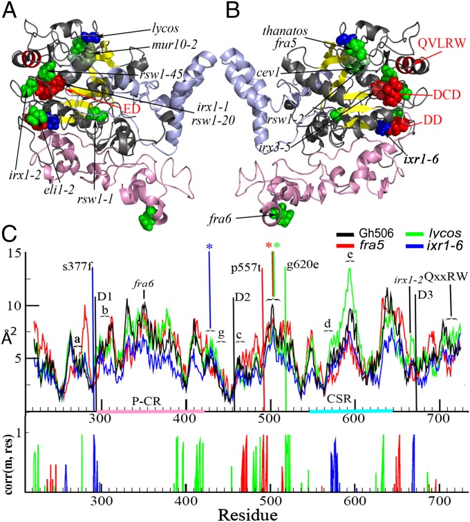
A 3D atomistic model of a plant cellulose synthase (CESA) has remained elusive despite over forty years of experimental effort. Here, we report a computationally predicted 3D structure of 506 amino acids of cotton CESA within the cytosolic region. Comparison of the predicted plant CESA structure with the solved structure of a bacterial cellulose-synthesizing protein validates the overall fold of the modeled glycosyltransferase (GT) domain. The coaligned plant and bacterial GT domains share a six-stranded β-sheet, five α-helices, and conserved motifs similar to those required for catalysis in other GT-2 glycosyltransferases. Extending beyond the cross-kingdom similarities related to cellulose polymerization, the predicted structure of cotton CESA reveals that plant-specific modules (plant-conserved region and class-specific region) fold into distinct subdomains on the periphery of the catalytic region. Computational results support the importance of the plant-conserved region and/or class-specific region in CESA oligomerization to form the multimeric cellulose-synthesis complexes that are characteristic of plants. Relatively high sequence conservation between plant CESAs allowed mapping of known mutations and two previously undescribed mutations that perturb cellulose synthesis in Arabidopsis thaliana to their analogous positions in the modeled structure. Most of these mutation sites are near the predicted catalytic region, and the confluence of other mutation sites supports the existence of previously undefined functional nodes within the catalytic core of CESA. Overall, the predicted tertiary structure provides a platform for the biochemical engineering of plant CESAs.


Conflict of interest statement

The authors declare no conflict of interest.

Figures

Fig. 1.
Fig. 1.
Predicted structure of the GhCESA cytosolic region. (A) Diagram of GhCESA1 showing eight predicted TMH and the large cytsolic loop between TMH2 and TMH3. Labels within the cytosolic loop indicate the approximate locations of the four conserved motifs; the P-CR region; the CSR region; and the analogous locations for published (black) and previously undescribed (red) missense mutations in Arabidopsis CESAs. (B) Snapshot of the Gh506 structure. The catalytic core is gray, the P-CR is pink, and the CSR is light blue. The catalytic core contains a β-sheet (yellow) with six strands: β-1 S287-S291; β-2 D253-S257; β-3 F454-D459; β-4 C532-N535; β-5 Y488-F491; and β-6 S686-C689. Green highlights DD, DCD, ED (directly behind DCD), and the QVLRW within α-13. The five α-helices that are part of the GT core are α-2 L267-A278; α-6 H433-V448; α-7 N466-D479; α-8 N508-K517; and α-13 S705-R725. (C) Diagram of the secondary structure showing six β-strands (yellow arrows) and 13 major α-helices (barrels) in three regions: catalytic core (red outlines); P-CR (pink fill); and CSR (blue fill). Possible additional shorter helical regions are indicated as unnumbered small barrels. (D) UDP-Glc (orange) docked into the catalytic site.
Fig. 2.
Fig. 2.
Possible oligomeric assemblies of the Gh506 cytosolic structure under (A) C2, (B) C3, (C) C4, and (D) C6 crystallization symmetries. The catalytic region is gray, the CSR is light blue, the P-CR is pink, QVLRW is yellow, and the site of fra6 mutations is red. (D) Bottom, (E) top, and (F) side view of the hexameric Gh506 assembly.
Fig. 3.
Fig. 3.
Comparison of the Gh506 with the bacterial cellulose synthase. (A) Surface representation of the Rs cellulose synthase (PDB ID 4HG6) superimposed with the Gh506 structure at the GT-domain (colored green). (B) Superimposition of the BcsA GT-domain with the Gh506 by secondary structure matching. The BcsA GT-domain is colored green, Gh506 GT-domain is gray and yellow, P-CR and CSR domains are colored pink and light blue, and UDP and glucan are shown as spheres. (C) Conserved sequence motifs that form the binding site for UDP and the acceptor glucan are compared between Gh506 (blue letters) and RsBcsA (black letters) with depicted residues in bold and colored as blue (Gh506) and gray (BcsA), and UDP (rust) and glucan (cyan).
Fig. 4.
Fig. 4.
Previously known (green) and previously undescribed (blue) Arabidopsis CESAs mutations mapped onto the Gh506 structure. (A and B) Two sides of Gh506 are shown with DD, DCD, ED, and QVLRW motifs in red. The equivalent GhCESA1 amino acid positions are: R351 (fra6); A436 (eli1-2); A447 (rsw1-1); D459 (irx3-5); P492 (fra5 and thanatos); G529 (rsw1-2); G531 (cev1) ; S668 (irx1-2); E671 (rsw1-45); D672 (irx1-1 and rsw1-20); and H680 (mur10-2). (C) Cross correlation of atomic fluctuations over simulation trajectories by residue. The peaks shown had at least 97% correlation, indicating distant effects of the mutations analogous to ixr1-6 (blue), lycos (green), and fra5 (red).

References

    1. Harris DM, et al. Cellulose microfibril crystallinity is reduced by mutating C-terminal transmembrane region residues CESA1A903V and CESA3T942I of cellulose synthase. Proc Natl Acad Sci USA. 2012;109(11):4098–4103. - PMC - PubMed
    1. Cantarel BL, et al. The Carbohydrate-Active EnZymes database (CAZy): An expert resource for Glycogenomics. Nucleic Acids Res. 2009;37(Database issue):D233–D238. - PMC - PubMed
    1. Somerville C. Cellulose synthesis in higher plants. Annu Rev Cell Dev Biol. 2006;22:53–78. - PubMed
    1. Nishiyama Y. Structure and properties of the cellulose microfi bril. J Wood Sci. 2009;55:241–249.
    1. Roberts E, Roberts AW. A cellulose synthase (Cesa) gene from the red alga Porphyra yezoensis (Rhodophyta) J Phycol. 2009;45:203–212. - PubMed

Publication types