. 2013 May 31;288(22):15699-711.
doi: 10.1074/jbc.M112.425066. Epub 2013 Apr 16.

Mutant copper-zinc superoxide dismutase (SOD1) induces protein secretion pathway alterations and exosome release in astrocytes: implications for disease spreading and motor neuron pathology in amyotrophic lateral sclerosis

Affiliations

Mutant copper-zinc superoxide dismutase (SOD1) induces protein secretion pathway alterations and exosome release in astrocytes: implications for disease spreading and motor neuron pathology in amyotrophic lateral sclerosis

Manuela Basso et al. J Biol Chem. .

Abstract

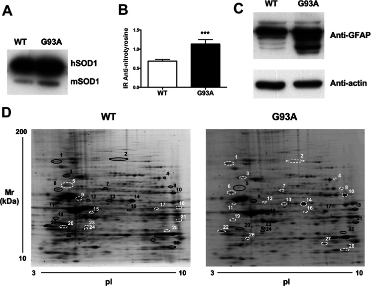
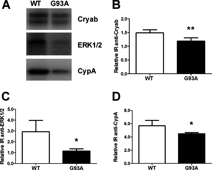
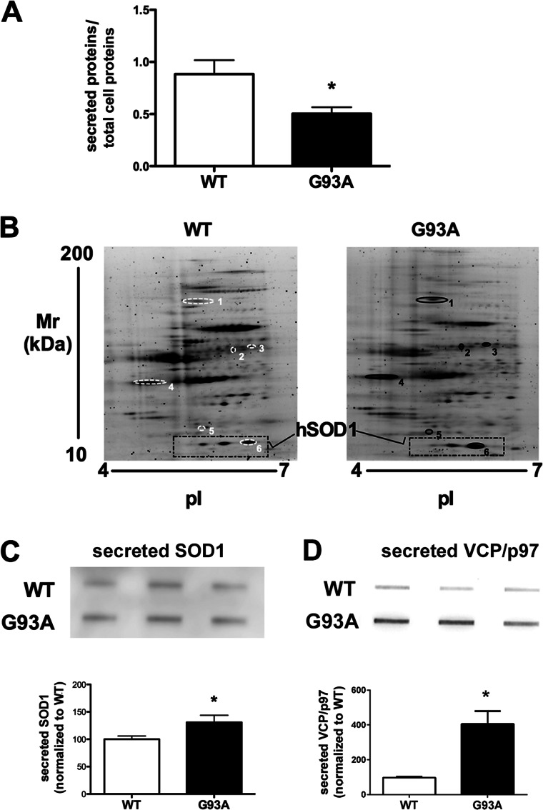
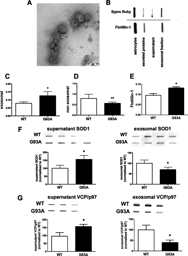
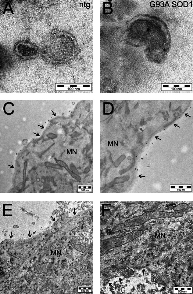
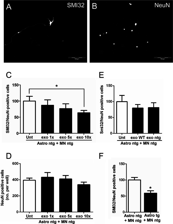
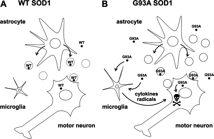
Amyotrophic lateral sclerosis is the most common motor neuron disease and is still incurable. The mechanisms leading to the selective motor neuron vulnerability are still not known. The interplay between motor neurons and astrocytes is crucial in the outcome of the disease. We show that mutant copper-zinc superoxide dismutase (SOD1) overexpression in primary astrocyte cultures is associated with decreased levels of proteins involved in secretory pathways. This is linked to a general reduction of total secreted proteins, except for specific enrichment in a number of proteins in the media, such as mutant SOD1 and valosin-containing protein (VCP)/p97. Because there was also an increase in exosome release, we can deduce that astrocytes expressing mutant SOD1 activate unconventional secretory pathways, possibly as a protective mechanism. This may help limit the formation of intracellular aggregates and overcome mutant SOD1 toxicity. We also found that astrocyte-derived exosomes efficiently transfer mutant SOD1 to spinal neurons and induce selective motor neuron death. We conclude that the expression of mutant SOD1 has a substantial impact on astrocyte protein secretion pathways, contributing to motor neuron pathology and disease spread.

Keywords: Amyotrophic Lateral Sclerosis (Lou Gehrig's Disease); Astrocytes; Disease Spreading; Exosomes; Proteomics; Superoxide Dismutase (SOD).


Figures

FIGURE 1.
FIGURE 1.
Characterization of primary astrocyte cultures derived from G93A and WT SOD1 mice and protein differential expression. A, cultures derived from G93A and WT SOD1 mice express similar amounts of human SOD1 (hSOD1) protein. A representative WB of human SOD1 levels and murine SOD1 (mSOD1) is also shown. B, slot blot for nitrotyrosine residues. Mutant SOD1 expression increases the levels of nitrotyrosine. Nitrotyrosine immunoreactivity (IR) was normalized to the total protein loaded, as measured after Coomassie staining. Values are the means ± S.E. (error bars) (n = 9). Asterisks indicate a G93A sample mean significantly higher (p < 0.001) than the WT sample mean, Student's t test. C, WB for GFAP in WT and G93A SOD1 astrocyte primary cultures. G93A SOD1 astrocytes present an increased number of GFAP fragments. D, 100 μg of proteins were loaded onto IPG strips (pI 3–10). Two-dimensional gel electrophoresis maps were stained with Sypro Ruby and analyzed with Progenesis software. The comparison revealed that the expression of mutant SOD1 is sufficient to alter the proteomic profile of the astrocytes (see Table 1). Black circles, up-regulated proteins; white circles, down-regulated proteins; spot numbers, proteins listed in Table 1.
FIGURE 2.
FIGURE 2.
Western blots of three differentially expressed proteins as validation of the proteomic analysis. A–D, representative WB for Cryab, ERK1/2, and CypA and quantification. Samples were 3-well pools of 6-well dishes from four separate primary astrocyte culture preparations. Ponceau Red staining was used as loading control. Values are relative immunoreactivities (IR) and are means ± S.E. (error bars) (n = 8). All of the proteins were significantly down-regulated (*, p < 0.05; **, p < 0.01) in astrocytes expressing mutant SOD1 in comparison with WT SOD1 controls (Student's t test).
FIGURE 3.
FIGURE 3.
G93A SOD1 astrocytes released a lower amount of proteins in the media. A, media were collected from similar numbers of cells expressing WT or G93A SOD1, and secreted proteins were prepared as described for slot blot experiments and quantified by BCA protein assay. Protein quantification was performed in at least seven samples per group. Each sample was a 3-well pool. Values are the amounts of secreted proteins normalized to total cell proteins (secreted proteins/total cell proteins) and are means ± S.E. (error bars). An asterisk indicates statistical significance (p < 0.05), Student's t test. B, for two-dimensional gel electrophoresis maps (n = 3 for each genotype), the same amount of secreted proteins (30 μg) were loaded on IPG strips (pI 4–7). The gel maps were stained with Sypro Ruby and compared by computerized image analysis. Six proteins were differentially secreted by astrocytes expressing mutant SOD1 and controls. Spot numbers refer to proteins listed in Table 2. C and D, slot blot and relative quantification of SOD1 (C) and VCP/p97 (D) released by the astrocytes confirmed the proteomic analysis and showed that the total level of the extracellular proteins were higher than in control conditions. The astrocytes were plated in 6-well dishes, and equal volumes of conditioned media from 3-well pools were used for the slot blot analysis. Values are immunoreactivities and are means ± S.E. (n = 3) normalized to WT, set as 100. An asterisk indicates statistical significance (p < 0.05), Student's t test.
FIGURE 4.
FIGURE 4.
WT and G93A SOD1 are released by the astrocytes, and different amounts are present in the exosomes. A, electron microscopy analysis of purified membrane vesicles with typical exosomal shape and dimension (diameter ranging from 80 to 140 nm; scale bar, 200 nm). B, slot blot of astrocyte protein lysate, secreted proteins, and ultracentrifuged proteins (supernatant and exosomal fractions). An exosomal marker, flotillin-1, is present in whole lysate, in unpurified secreted proteins, and in the exosomal fraction but not in the supernatant. C and E, G93A SOD1 expression increases exosomal proteins. Protein quantification of the supernatant and exosomal fractions was done by BCA protein assay in at least seven samples per genotype. Each sample was a 3-well pool. Values are the amounts of non-exosomal or exosomal proteins normalized to total astrocytic proteins (non-exosomal or exosomal proteins/total astrocytic proteins) and are means ± S.E. (error bars) Asterisks indicate statistical significance (*, p < 0.05; **, p < 0.01), Student's t test. D, flotillin-1 was measured by slot blot in WT and G93A SOD1 astrocyte-conditioned media. Anti-flotillin-1 immunoreactivity was normalized to the total protein loaded, as measured after Sypro Ruby blot staining. Values are means ± S.E. (n = 3). The asterisk indicates a G93A sample mean significantly higher (p < 0.05) than the WT sample mean, Student's t test. F and G, slot blot for SOD1 and VCP/p97 in the exosomal and non-exosomal (supernatant) fractions. Equal volumes of exosomal or non-exosomal fractions were used to measure protein levels in the G93A SOD1 and WT SOD1 conditions. Each sample was taken from a 3-well pool. Values are immunoreactivities and are means ± S.E. (n = 4) normalized to WT, set as 100. An asterisk indicates statistical significance (p < 0.05), Student's t test.
FIGURE 5.
FIGURE 5.
Mutant SOD1 is transferred to spinal neurons through astrocyte-derived exosomes. Electron microscopy pictures of exosomes with characteristic cup-shaped morphology from non-transgenic (ntg) (A) and G93A SOD1-expressing (B) astrocytes (scale bar, 100 nm). Immunoelectron microscopy using anti-human SOD1 antibody indicated SOD1 exclusively inside exosomes isolated from transgenic human SOD1-overexpressing astrocytes (in this case G93A SOD1; see also supplemental Fig. S1A for WT SOD1). C and D, two representative images at different magnification (scale bar, 500 nm) of cultured non-transgenic spinal neurons exposed to G93A SOD1-containing exosome-depleted fractions, showing the 10-nm electron-dense round gold particles labeling human SOD1 only outside the cell; treatment with G93A SOD1-containing exosomes (E and F) caused the diffusion inside the cytoplasm of the majority of human SOD1 (scale bar, 500 nm). The same results were obtained with WT SOD1-containing exosomes (supplemental Fig. S1B). The arrows indicate the plasma membrane of a motor neuron (MN).
FIGURE 6.
FIGURE 6.
G93A SOD1 astrocyte-derived exosomes are sufficient to induce motor neuron death. In spinal neuron-astrocyte co-cultures, motor neuron survival was determined by the ratio of the number of SMI32-positive cells (A) to total NeuN-positive cells (B) in the same frame (20 frames/well). Scale bar, 100 μm. C, non-transgenic motor neurons plated on non-transgenic astrocytes (Astro ntg + MN ntg) survived significantly less after 6 days in culture when treated with increasing concentrations (1×, 5×, and 10×) of G93A SOD1 astrocyte-derived exosomes (post-test for linear trend p = 0.03; n = 8 for each condition). The 10× exosome concentration (exo 10×) significantly reduced the survival of motor neurons compared with untreated (Unt) (Student's t test, p = 0.01). D, neurons (NeuN-positive cells) did not show a significantly reduced survival, compared with untreated, by post-test linear trend (p = 0.27), even with the 10× exosome concentration (Student's t test, p > 0.05). Values are means of NeuN-positive cells per well. E, non-transgenic motor neurons plated on non-transgenic astrocytes showed no significantly reduced survival after 6 days in culture when treated with a 10× concentration of WT SOD1 (exo WT) or non-transgenic (exo ntg) astrocyte-derived exosomes with respect to untreated cells, Student's t test (n = 8 for each condition). F, a toxic effect was observed when non-transgenic spinal neurons were plated on G93A SOD1 astrocytes (Astro tg + MN ntg) after 6 days in culture (Student's t test, p = 0.0003; n = 17 for each condition). Error bars, S.E.
FIGURE 7.
FIGURE 7.
Proposed pathogenic mechanism for G93A SOD1-expressing astrocytes. A, astrocytes normally release WT SOD1 in the extracellular space through exosomal and non-exosomal secretory pathways. B, astrocytes expressing mutant SOD1 (G93A SOD1) activate unconventional secretory pathways, possibly to protect themselves from mutant SOD1-associated toxicity and release more exosomes. Exosomes contribute to the spreading of the disease by continuous transfer of toxic molecules, including G93A SOD1, to neighboring motor neurons. G93A SOD1 secreted by astrocytes contributes to motor neuron injury indirectly, increasing the release of proinflammatory cytokines and free radicals by activated microglia (17, 62) and directly, once it is transferred to inside motor neurons by exosomes, by facilitating the initiation of aggregation.

References

    1. Ilieva H., Polymenidou M., Cleveland D. W. (2009) Non-cell autonomous toxicity in neurodegenerative disorders. ALS and beyond. J. Cell Biol. 187, 761–772 - PMC - PubMed
    1. Howland D. S., Liu J., She Y., Goad B., Maragakis N. J., Kim B., Erickson J., Kulik J., DeVito L., Psaltis G., DeGennaro L. J., Cleveland D. W., Rothstein J. D. (2002) Focal loss of the glutamate transporter EAAT2 in a transgenic rat model of SOD1 mutant-mediated amyotrophic lateral sclerosis (ALS). Proc. Natl. Acad. Sci. U.S.A. 99, 1604–1609 - PMC - PubMed
    1. Rothstein J. D., Van Kammen M., Levey A. I., Martin L. J., Kuncl R. W. (1995) Selective loss of glial glutamate transporter GLT-1 in amyotrophic lateral sclerosis. Ann. Neurol. 38, 73–84 - PubMed
    1. Bendotti C., Tortarolo M., Suchak S. K., Calvaresi N., Carvelli L., Bastone A., Rizzi M., Rattray M., Mennini T. (2001) Transgenic SOD1 G93A mice develop reduced GLT-1 in spinal cord without alterations in cerebrospinal fluid glutamate levels. J. Neurochem. 79, 737–746 - PubMed
    1. Aebischer J., Cassina P., Otsmane B., Moumen A., Seilhean D., Meininger V., Barbeito L., Pettmann B., Raoul C. (2011) IFNγ triggers a LIGHT-dependent selective death of motoneurons contributing to the non-cell-autonomous effects of mutant SOD1. Cell Death Differ. 18, 754–768 - PMC - PubMed

Publication types

MeSH terms