

Erdheim-Chester disease with isolated craniocerebral involvement
- PMID: 23592809
- PMCID: PMC3645836
- DOI: 10.1136/bcr-2012-006823
Erdheim-Chester disease with isolated craniocerebral involvement
Abstract
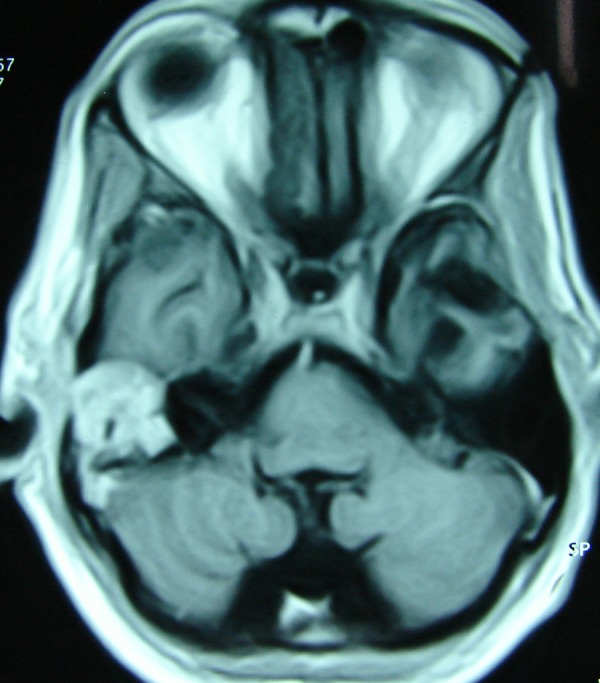
Erdheim-Chester disease (ECD) is a rare non-Langerhans form of histiocytosis with distinctive radiographic and pathological features. Intracranial involvement is further a rarity, usually producing diabetes insipidus or cerebellar-brainstem symptoms. We report a 40-year-old man presenting with recurrent secondarily generalised seizures. An MRI scan of the brain revealed multiple enhancing intracranial masses in frontal, temporal and parietal regions. Biopsy from the left frontotemporal lesion confirmed it to be a rare case of ECD. The patient received a short course of corticosteroids initially and subsequently remained well-controlled on antiepileptic therapy alone. A repeat MRI of his brain showed significant resolution of lesions. Osteolytic lesions in the skull vault were detected during follow-up which also disappeared. Interestingly, there was no involvement of long bones or any other system even after 12 years of follow-up.
Figures


References
-
- Drier A, Haroche J, Savotovsky J, et al. Cerebral, facial, and orbital involvement in Erdheim-Chester disease: CT and MR findings. Radiology 2010;2013:586–94 - PubMed
-
- Bianco F, Lacovelli E, Tinelli E, et al. Characteristic brain MRI appearance of Erdheim-Chester disease. Neurology 2009;2013:2120–2 - PubMed
-
- Adem C, Helie O, Leveque C, et al. Case 78: Erdheim-Chester disease with central nervous system involvement. Radiology 2005;2013:111–15 - PubMed
-
- Adle-Biassette H, Chetritt J, Bergemer-Fouquet AM, et al. Pathology of the central nervous system in Chester-Erdheim disease: report of three cases. J Neuropathol Exp Neurol 1997;2013:1207–16 - PubMed
-
- Veyssier-Belot C, Cacoub P, Caparros-Lefebvre D, et al. Erdheim-Chester disease: clinical and radiologic characteristics of 59 cases. Medicine 1996;2013:157–69 - PubMed
Publication types
MeSH terms
Substances
LinkOut - more resources
Full Text Sources
Other Literature Sources
Medical