

Case Reports
doi: 10.1136/bcr-2012-008515.
Fetal supraventricular tachycardia, treating the baby by targeting the mother
Affiliations
- PMID: 23592812
- PMCID: PMC3644937
- DOI: 10.1136/bcr-2012-008515
Item in Clipboard
Case Reports
Fetal supraventricular tachycardia, treating the baby by targeting the mother
BMJ Case Rep.
.
Abstract
Fetal supraventricular tachycardia (SVT) is the most common form of fetal tachycardia. If started early in pregnancy, it can cause non-immune fetal hydrops. Echocardiography is the preferred method for the diagnosis with simultaneous pulsed Doppler recording from the superior vena cava and ascending aorta. Transplacental therapy with digoxin is the most common way of treatment. We present a case of fetal SVT detected at 26 weeks of pregnancy. Digoxin therapy restored the rhythm initially, but later paroxysms of fetal SVT persisted necessitating the addition of second antiarrhythmic medication which was discussed with the parents. The couple chose to proceed for premature delivery at 32 weeks.
Figures
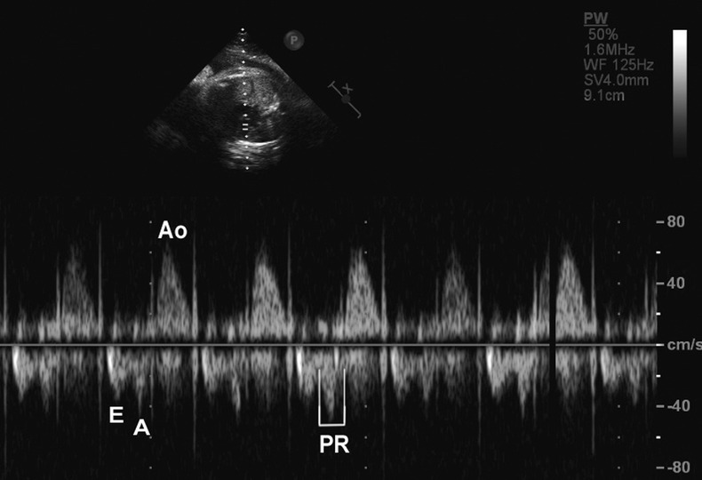
Fetal echocardiography still image of simultaneous mitral inflow and aortic outflow obtained via five-chamber view. The heart rate was 143/min. The positive signal is due to aortic outflow and the negative signals are due to mitral inflow. The mechanical PR interval was 95 m/s. Ao, aortic outflow; E, mitral E wave; A, mitral A wave.
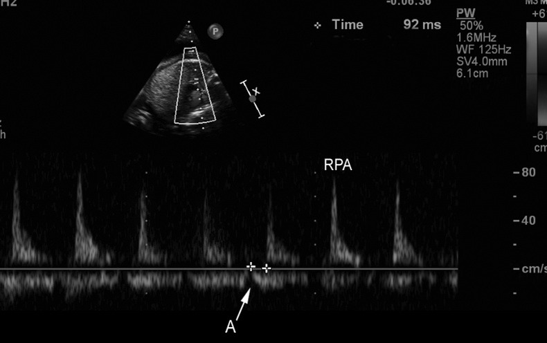
Simultaneous pulse Doppler recording during sinus rhythm, from the right pulmonary artery (positive signal) and right pulmonary vein (negative signal). The atrial contraction is denoted by an indentation in the pulmonary vein signal. The mechanical PR interval in this recording was 92 m/s.
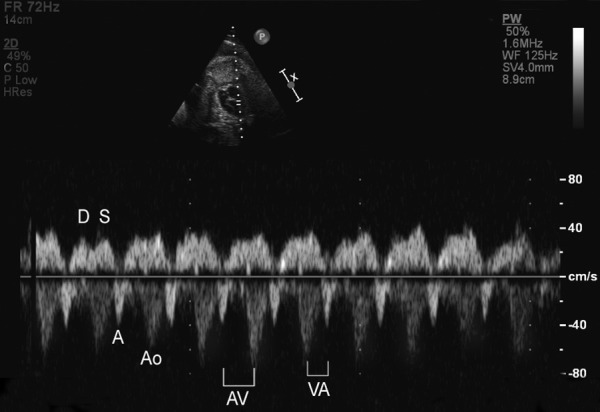
Simultaneous pulse Doppler recording from the superior vena cava (SVC) and ascending aorta. The positive signal is caused by SVC blood flow, while the negative signal is caused by aortic flow. The A wave reversal is seen as negative signal, moving away from the heart. Note that the AV interval is longer than the VA interval. Ao, aortic outflow; D, SVC D wave (diastole); S, SVC S wave (systole); A, SVC A wave reversal (atrial contraction).
Similar articles
-
Amiodarone in treatment of fetal supraventricular tachycardia. A case report and review of literature.Fetal Diagn Ther. 2006;21(1):72-6. doi: 10.1159/000089052. Fetal Diagn Ther. 2006. PMID: 16354980 Review.
-
Fetal supraventricular tachycardia complicated by hydrops fetalis: a role for direct fetal intramuscular therapy.Am J Perinatol. 1996 Nov;13(8):483-6. doi: 10.1055/s-2007-994432. Am J Perinatol. 1996. PMID: 8989479
-
Successful digoxin therapy of fetal supraventricular tachycardia in a triplet pregnancy.Obstet Gynecol. 2001 Nov;98(5 Pt 2):921-3. doi: 10.1016/s0029-7844(01)01436-3. Obstet Gynecol. 2001. PMID: 11704204 Review.
-
Antenatal Therapy for Fetal Supraventricular Tachyarrhythmias: Multicenter Trial.J Am Coll Cardiol. 2019 Aug 20;74(7):874-885. doi: 10.1016/j.jacc.2019.06.024. J Am Coll Cardiol. 2019. PMID: 31416531 Clinical Trial.
-
Refractory supraventricular fetal tachycardia as a cause of non-immune hydrops: management conundrum.BMJ Case Rep. 2023 Dec 28;16(12):e255808. doi: 10.1136/bcr-2023-255808. BMJ Case Rep. 2023. PMID: 38154874
Cited by
-
Fetal supraventricular tachycardia and maternal COVID-19 vaccination: is there any relationship?Future Sci OA. 2022 Sep 12;8(7):FSO812. doi: 10.2144/fsoa-2022-0007. eCollection 2022 Aug. Future Sci OA. 2022. PMID: 36248062 Free PMC article.
-
Successful medical treatment of fetal supraventricular tachycardia that cause hydrops fetalis.Turk J Obstet Gynecol. 2014 Sep;11(3):193-195. doi: 10.4274/tjod.56578. Epub 2014 Sep 15. Turk J Obstet Gynecol. 2014. PMID: 28913017 Free PMC article.
-
Maternal monitoring and safety considerations during antiarrhythmic treatment for fetal supraventricular tachycardia.Obstet Med. 2019 Jun;12(2):66-75. doi: 10.1177/1753495X18808118. Epub 2018 Nov 15. Obstet Med. 2019. PMID: 31217810 Free PMC article. Review.
-
Morphine-induced supraventricular tachycardia in near-term fetus.Ital J Pediatr. 2018 Sep 24;44(1):111. doi: 10.1186/s13052-018-0570-1. Ital J Pediatr. 2018. PMID: 30249290 Free PMC article.
-
Unexpected Rhythm: Supraventricular Tachycardia Unveiled in a Neonate Diagnosed at Delivery.Cureus. 2024 Jul 29;16(7):e65710. doi: 10.7759/cureus.65710. eCollection 2024 Jul. Cureus. 2024. PMID: 39211675 Free PMC article.
References
-
- Hyman AS. Irregularities of the fetal heart: a phonocardiographic study of the fetal heart sounds from the fifth to eighth months of pregnancy. Am J Obstet Gynecol 1930;2013:332–47
-
- Silber DL, Durnin RE. Intrauterine atrial tachycardia, associated with massive edema in a newborn. Am J Dis Child 1969;2013:722–6 - PubMed
-
- Perles Z, Gavri S, Rein AJJT, et al. Tachyarrhythmias in the fetus: state of the art diagnosis and treatment. Prog Pediatr Cardiol 2006;2013:95–107
-
- Silver LE, Platt LD, Santulli TV, Jr, et al. Resolution of hydrops fetalis despite persistent fetal tachycardia. J Ultrasound Med 2001;2013:1141–5 - PubMed
-
- Rana YS, Sodhi B, Kochar SPS, et al. Successful digoxin therapy of fetal supraventricular tachycardia. S Asian Fed Obstet Gynecol 2009;2013:44–6
Publication types
MeSH terms
Substances
LinkOut - more resources
Full Text Sources
Other Literature Sources
Medical