The JAK-STAT transcriptional regulator, STAT-5, activates the ATM DNA damage pathway to induce HPV 31 genome amplification upon epithelial differentiation
- PMID: 23593005
- PMCID: PMC3616964
- DOI: 10.1371/journal.ppat.1003295
The JAK-STAT transcriptional regulator, STAT-5, activates the ATM DNA damage pathway to induce HPV 31 genome amplification upon epithelial differentiation
Abstract
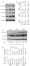
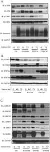
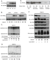
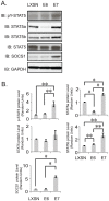
High-risk human papillomavirus (HPV) must evade innate immune surveillance to establish persistent infections and to amplify viral genomes upon differentiation. Members of the JAK-STAT family are important regulators of the innate immune response and HPV proteins downregulate expression of STAT-1 to allow for stable maintenance of viral episomes. STAT-5 is another member of this pathway that modulates the inflammatory response and plays an important role in controlling cell cycle progression in response to cytokines and growth factors. Our studies show that HPV E7 activates STAT-5 phosphorylation without altering total protein levels. Inhibition of STAT-5 phosphorylation by the drug pimozide abolishes viral genome amplification and late gene expression in differentiating keratinocytes. In contrast, treatment of undifferentiated cells that stably maintain episomes has no effect on viral replication. Knockdown studies show that the STAT-5β isoform is mainly responsible for this activity and that this is mediated through the ATM DNA damage response. A downstream target of STAT-5, the peroxisome proliferator-activated receptor γ (PPARγ) contributes to the effects on members of the ATM pathway. Overall, these findings identify an important new regulatory mechanism by which the innate immune regulator, STAT-5, promotes HPV viral replication through activation of the ATM DNA damage response.
Conflict of interest statement
The authors have declared that no competing interests exist.
Figures


Similar articles
-
The acetyltransferase Tip60 is a critical regulator of the differentiation-dependent amplification of human papillomaviruses.J Virol. 2015 Apr;89(8):4668-75. doi: 10.1128/JVI.03455-14. Epub 2015 Feb 11. J Virol. 2015. PMID: 25673709 Free PMC article.
-
STAT-5 Regulates Transcription of the Topoisomerase IIβ-Binding Protein 1 (TopBP1) Gene To Activate the ATR Pathway and Promote Human Papillomavirus Replication.mBio. 2015 Dec 22;6(6):e02006-15. doi: 10.1128/mBio.02006-15. mBio. 2015. PMID: 26695634 Free PMC article.
-
Human papillomaviruses activate the ATM DNA damage pathway for viral genome amplification upon differentiation.PLoS Pathog. 2009 Oct;5(10):e1000605. doi: 10.1371/journal.ppat.1000605. Epub 2009 Oct 2. PLoS Pathog. 2009. PMID: 19798429 Free PMC article.
-
DNA damage response is hijacked by human papillomaviruses to complete their life cycle.J Zhejiang Univ Sci B. 2017 Mar;18(3):215-232. doi: 10.1631/jzus.B1600306. J Zhejiang Univ Sci B. 2017. PMID: 28271657 Free PMC article. Review.
-
Manipulation of the innate immune response by human papillomaviruses.Virus Res. 2017 Mar 2;231:34-40. doi: 10.1016/j.virusres.2016.11.004. Epub 2016 Nov 5. Virus Res. 2017. PMID: 27826042 Free PMC article. Review.
Cited by
-
Activating the DNA Damage Response and Suppressing Innate Immunity: Human Papillomaviruses Walk the Line.Pathogens. 2020 Jun 13;9(6):467. doi: 10.3390/pathogens9060467. Pathogens. 2020. PMID: 32545729 Free PMC article. Review.
-
KLF13 regulates the differentiation-dependent human papillomavirus life cycle in keratinocytes through STAT5 and IL-8.Oncogene. 2016 Oct 20;35(42):5565-5575. doi: 10.1038/onc.2016.97. Epub 2016 Apr 4. Oncogene. 2016. PMID: 27041562
-
SAMHD1 Regulates Human Papillomavirus 16-Induced Cell Proliferation and Viral Replication during Differentiation of Keratinocytes.mSphere. 2019 Aug 7;4(4):e00448-19. doi: 10.1128/mSphere.00448-19. mSphere. 2019. PMID: 31391281 Free PMC article.
-
The molecular biology and HPV drug responsiveness of cynomolgus macaque papillomaviruses support their use in the development of a relevant in vivo model for antiviral drug testing.PLoS One. 2019 Jan 25;14(1):e0211235. doi: 10.1371/journal.pone.0211235. eCollection 2019. PLoS One. 2019. PMID: 30682126 Free PMC article.
-
Productive replication of human papillomavirus 31 requires DNA repair factor Nbs1.J Virol. 2014 Aug;88(15):8528-44. doi: 10.1128/JVI.00517-14. Epub 2014 May 21. J Virol. 2014. PMID: 24850735 Free PMC article.
References
-
- zur Hausen H (2002) Papillomaviruses and cancer: from basic studies to clinical application. Nat Rev Cancer 2: 342–350. - PubMed
-
- Moody CA, Laimins LA (2010) Human papillomavirus oncoproteins: pathways to transformation. Nat Rev Cancer 10: 550–560. - PubMed
-
- Munger K, Howley PM (2002) Human papillomavirus immortalization and transformation functions. Virus Res 89: 213–228. - PubMed
-
- Scheffner M, Werness BA, Huibregtse JM, Levine AJ, Howley PM (1990) The E6 oncoprotein encoded by human papillomavirus types 16 and 18 promotes the degradation of p53. Cell 63: 1129–1136. - PubMed
Publication types
MeSH terms
Substances
Grants and funding
LinkOut - more resources
Full Text Sources
Other Literature Sources
Research Materials
Miscellaneous

