. 2013 Apr;9(4):e1003310.
doi: 10.1371/journal.pgen.1003310. Epub 2013 Apr 4.

Site-specific phosphorylation of the DNA damage response mediator rad9 by cyclin-dependent kinases regulates activation of checkpoint kinase 1

Affiliations

Site-specific phosphorylation of the DNA damage response mediator rad9 by cyclin-dependent kinases regulates activation of checkpoint kinase 1

Carla Manuela Abreu et al. PLoS Genet. 2013 Apr.

Abstract

The mediators of the DNA damage response (DDR) are highly phosphorylated by kinases that control cell proliferation, but little is known about the role of this regulation. Here we show that cell cycle phosphorylation of the prototypical DDR mediator Saccharomyces cerevisiae Rad9 depends on cyclin-dependent kinase (CDK) complexes. We find that a specific G2/M form of Cdc28 can phosphorylate in vitro the N-terminal region of Rad9 on nine consensus CDK phosphorylation sites. We show that the integrity of CDK consensus sites and the activity of Cdc28 are required for both the activation of the Chk1 checkpoint kinase and its interaction with Rad9. We have identified T125 and T143 as important residues in Rad9 for this Rad9/Chk1 interaction. Phosphorylation of T143 is the most important feature promoting Rad9/Chk1 interaction, while the much more abundant phosphorylation of the neighbouring T125 residue impedes the Rad9/Chk1 interaction. We suggest a novel model for Chk1 activation where Cdc28 regulates the constitutive interaction of Rad9 and Chk1. The Rad9/Chk1 complex is then recruited at sites of DNA damage where activation of Chk1 requires additional DDR-specific protein kinases.


Conflict of interest statement

The authors have declared that no competing interests exist.

Figures

Figure 1
Figure 1. Rad9 is phosphorylated by Cdc28 in the absence of DNA damage.
(A) Cell cycle regulation of phosphorylated forms of Rad9. An α-factor block and release experiment using yeast strain CG378. Rad9 phospho-forms were identified in protein extracts by western blotting. The phospho-protein Swi6 serves as a loading control. The cell cycle phases of synchronously cycling cells determined by budding analysis are indicated above the blots. The insert in the graph shows Rad9 from asynchronously growing cells or cells arrested with α-factor, HU or nocodazole. (B) Rad9 phosphorylation is dependent on Cdc28 activity. Rad9 and Orc6 western blots performed with extracts prepared from asynchronously growing, α-factor (G1) or nocodazole (G2/M) arrested cdc28-as1 cells either mock or 1-NMPP1 treated. (C) Rad9 phosphorylation is dependent on Cdc28/Clb activity. The indicated cells were arrested with α-factor, shifted to 30°C and samples were taken at the indicated times for Rad9 western and FACS analysis. See also Figure S1 for related experiments.
Figure 2
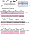
Figure 2. N-terminal CDK consensus sites are phosphorylated by Cdc28/Clb2 and are required for activation of Chk1 in vivo.
(A) Schematic representation of the Rad9 N-terminus showing the 9 consensus sites for phosphorylation by Cdc28 and those mutated to alanine in Rad9CDK1-9A mutant protein. (B) In vitro phosphorylation of Rad9 CADWT, but not CADCDK1-9A, requires Cdc28/Clb2. In vitro kinase assays were performed on the indicated substrates with four specific purified Cdc28 complexes. (C) DNA damage-dependent Chk1 phosphorylation is defective in rad9CDK1-9A, rad9CADΔ and rad9 Δ cells. Asynchronously growing cells were treated with bleocin for the indicated times and Chk1 phosphorylation analysed by western blotting. (D&E) Chk1 activation is mostly dependent on the 9 CAD CDK consensus sites in G2/M cells. Cells were grown and arrested in the cell cycle as indicated with either α-factor (G1 cells in D) or nocodazole (G2/M cells in E), treated with bleocin for the indicated times and analysed by western blotting of Chk1-3HA (D & E). See also Figure S2 for related experiments.
Figure 3
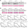
Figure 3. The CDK1-9 sites of Rad9 specifically function in the Chk1 branch of the G2/M checkpoint.
rad9CDK1-9A mutant cells arrested in G2/M by nocodazole treatment are neither defective for Rad9 (A) nor for Rad53 (B) phosphorylations induced by bleocin treatment started at time 0. (C) The CDK1-9 sites of Rad9 function specifically in the Chk1 branch and not the Rad53 branch of the G2/M checkpoint. The indicated strains were examined for epistatic relationships using the G2/M checkpoint assay, in which cells synchronized in G2/M using nocodazole are released after irradiation into medium without nocodazole, but containing α-factor, preventing cells that have successfully completed mitosis from cycling further by arresting them in G1. This assay measures the delay in completing mitosis under DNA damaging conditions by comparing the behavior of cells that have been irradiated with IR (+IR) or not (−IR). All strains contained the sml1Δ mutation necessary for the viability of rad53Δ cells. See also Figure S3.
Figure 4
Figure 4. CDK is required for the activation of Chk1-dependent signaling.
Cdc28 activity was regulated using the 1-NMPP1 inhibitor in G2/M arrested cdc28-as1 cells treated with bleocin or 4-NQO to examine the activation of Chk1 signaling. Rad9 and Rad53 were followed as markers of checkpoint activation, while Orc6 phosphorylation serves as a marker for Cdc28 inactivation. See also Figure S4.
Figure 5
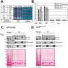
Figure 5. CDK-dependent phosphorylation of the nine N-terminal CDK sites in Rad9 regulates a physical interaction between Rad9 and Chk1.
(A) Rad9/Chk1 interaction measured in vivo using a yeast two-hybrid (Y2H) assay is dependent on the CDK1-9 sites. Y2H interaction of specific bait and prey plasmids shown on the left is indicated by the white colour of the otherwise red cells, their resistance to Aureobasidin A and their blue colour on media containing the X-α-gal substrate, as for the p53/T antigen interaction control. Six independent clones are presented for each vector combination. (B) The Y2H interaction between Rad9 and Chk1 is dependent on CDK activity in G2/M cells. The indicated bait and prey plasmids were introduced into cdc28-as1 cells mock treated or treated with 1-NMPP1 1 h after synchronization of cells with either nocodazole or alpha factor and prior to induction of expression of each bait protein. The recently reported CDK-dependent Rad9/Dpb11 interaction was used as a positive control. (C) The Rad9/Chk1 interaction measured using co-immunoprecipitation (co-IP) occurs both in the absence and presence of DNA damage. Chk1 (anti-FLAG) and Dpb11 (anti-MYC) immunoprecipitations (IPs) were performed as indicated on extracts prepared from nocodazole-arrested cells, expressing both Chk1-3FLAG and Dpb11-13MYC, and either mock treated or treated with 20 µg/ml of bleocin for 45 min. Mock (IgG) or Dpb11 (MYC) IPs were performed as controls. Rad9, Chk1-3FLAG and Dpb11-13MYC specific bands were detected in western blots. Lower exposures, to facilitate their visualisation, of the western blots of the starting extracts are shown to the left. (D) Chk1 interaction with Rad9 is dependent on the CDK1-9 sites. As in panel C, except rad9CDK1-9A cells were used. See also Figure S5 for related experiments.
Figure 6
Figure 6. CDK sites 6 (T125) and 7 (T143) regulate the Rad9/Chk1 interaction.
(A) The Y2H interaction between Rad9 and Chk1 mostly requires the 6th and 7th CDK sites, T125 and T143, of the CAD region (B) Peptides pull down experiments identified phosphorylated T143 (CDK site 7) and unphosphorylated T125 (CDK site 6) as the best combination of these sites required for maximum Chk1/Rad9 interaction. Chk1-3FLAG was immunopurified from rad9Δ cell clarified crude extracts (CCE) and incubated with four different biotinylated 35 amino-acid peptides. As represented in red, the T125 T143, T125p, T143p and T125p143p peptides were un-, mono- or di-phosphorylated on residues T125 and T143. Magnetic streptavidin beads were boiled to analyse both the presence of Chk1-3FLAG (anti-FLAG western blot) and that equal amount of peptides were used in each pull down (see Ponceau S stain). The dependency on the phosphorylation of T143 was confirmed by lambda phosphatase treatment in the absence (λ) or in the presence of phosphatase inhibitors (λ+Inh). Empty lanes are indicated by ‘x’ above the relevant lanes. (C) The Rad9 CAD peptide array phosphorylation profile shows that peptides containing consensus CDK sites 6 (T125), 7 (T143), 8 (T155) and 9 (T218) are phosphorylated by Cdc28/Clb2 in vitro. Peptides arrays of immobilized overlapping 19-mer peptides, each shifted to the right by 3 amino acids encompassing the first 260 amino acids of Rad9 sequence, were generated and are schematically represented at the top of the panel. The arrays were used in a kinase assay with (+) or without (−) the purified Cdc28/Clb2 complex. Target peptides of Cdc28, CK2 and Cdc7 were also spotted as controls as indicated. (D) Phosphorylation levels for CDK site-containing peptides within Rad9 isolated from asynchronous, G1- and G2/M arrested cells. Relative abundances were determined by mass spectrometry (see Material and Methods section). Residues T125 and T143 (CDK sites 6 and 7 respectively) are differentially phosphorylated in vivo. ○ indicates phosphorylated peptide containing this CDK site is assigned by HPLC retention time and accurate mass only. ND = Not detected. Non-phosphorylated and phosphorylated peptides were not detected. See also Figure S7, Table S1 and Table S5.
Figure 7
Figure 7. Model of Chk1 activation in response to DNA damage.
Note that the tetramer representation of Rad9 is a possible conformation for the C-Rad9 oligomeric complex of greater than or equal to 850 kDa, which is remodelled into a smaller complex of approximately 560 kDa containing D-Rad9 after DNA damage. See text for a description of the model.

References

    1. Harper JW, Elledge SJ (2007) The DNA damage response: ten years after. Mol Cell 28: 739–745. - PubMed
    1. Halazonetis TD, Gorgoulis VG, Bartek J (2008) An oncogene-induced DNA damage model for cancer development. Science 319: 1352–1355. - PubMed
    1. Durocher D, Jackson SP (2001) DNA-PK, ATM and ATR as sensors of DNA damage: variations on a theme? Curr Opin Cell Biol 13: 225–231. - PubMed
    1. Stracker TH, Usui T, Petrini JH (2009) Taking the time to make important decisions: the checkpoint effector kinases Chk1 and Chk2 and the DNA damage response. DNA Repair (Amst) 8: 1047–1054. - PMC - PubMed
    1. FitzGerald JE, Grenon M, Lowndes NF (2009) 53BP1: function and mechanisms of focal recruitment. Biochem Soc Trans 37: 897–904. - PubMed

Publication types

MeSH terms