. 2013 Apr;9(4):e1003412.
doi: 10.1371/journal.pgen.1003412. Epub 2013 Apr 4.

Drosophila DJ-1 decreases neural sensitivity to stress by negatively regulating Daxx-like protein through dFOXO

Affiliations

Drosophila DJ-1 decreases neural sensitivity to stress by negatively regulating Daxx-like protein through dFOXO

Soojin Hwang et al. PLoS Genet. 2013 Apr.

Abstract

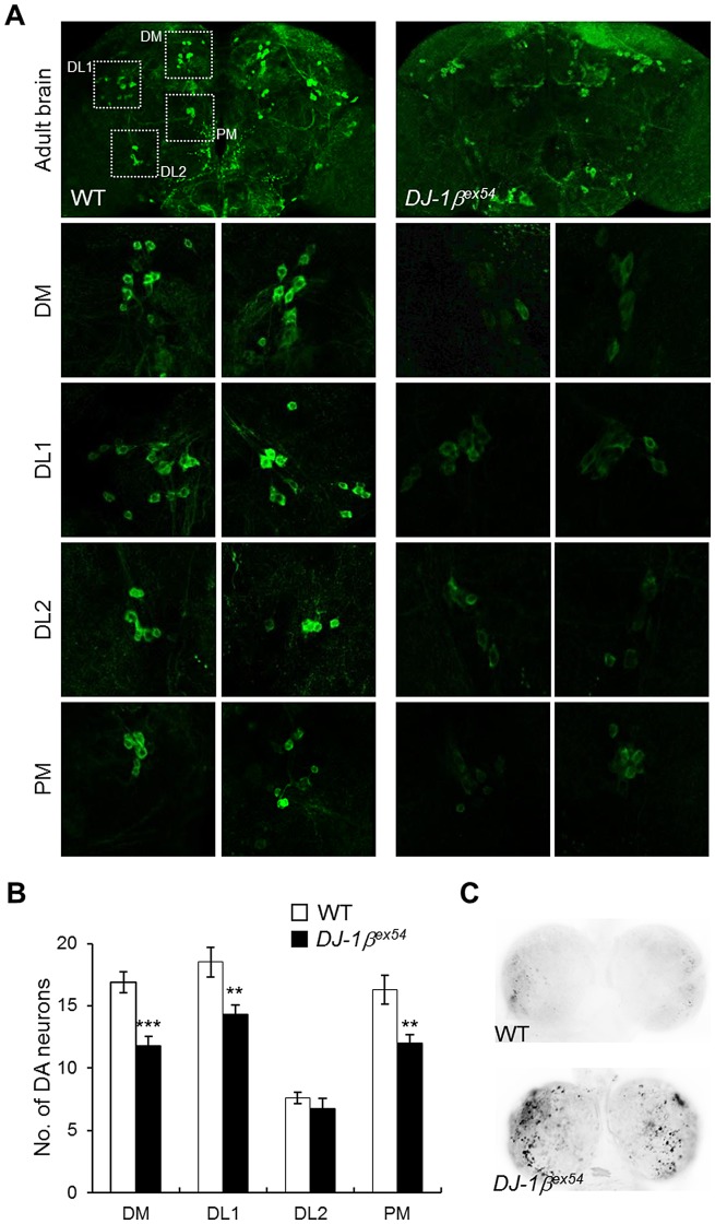
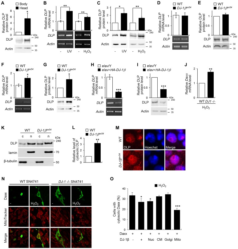
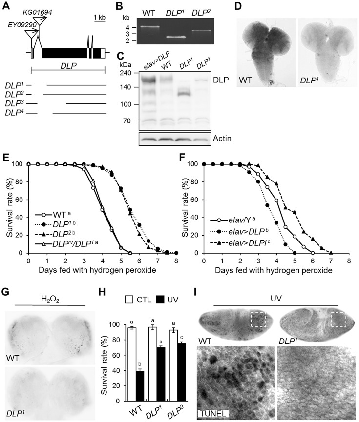
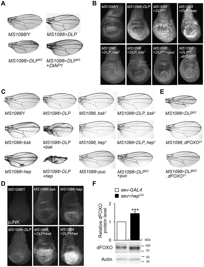
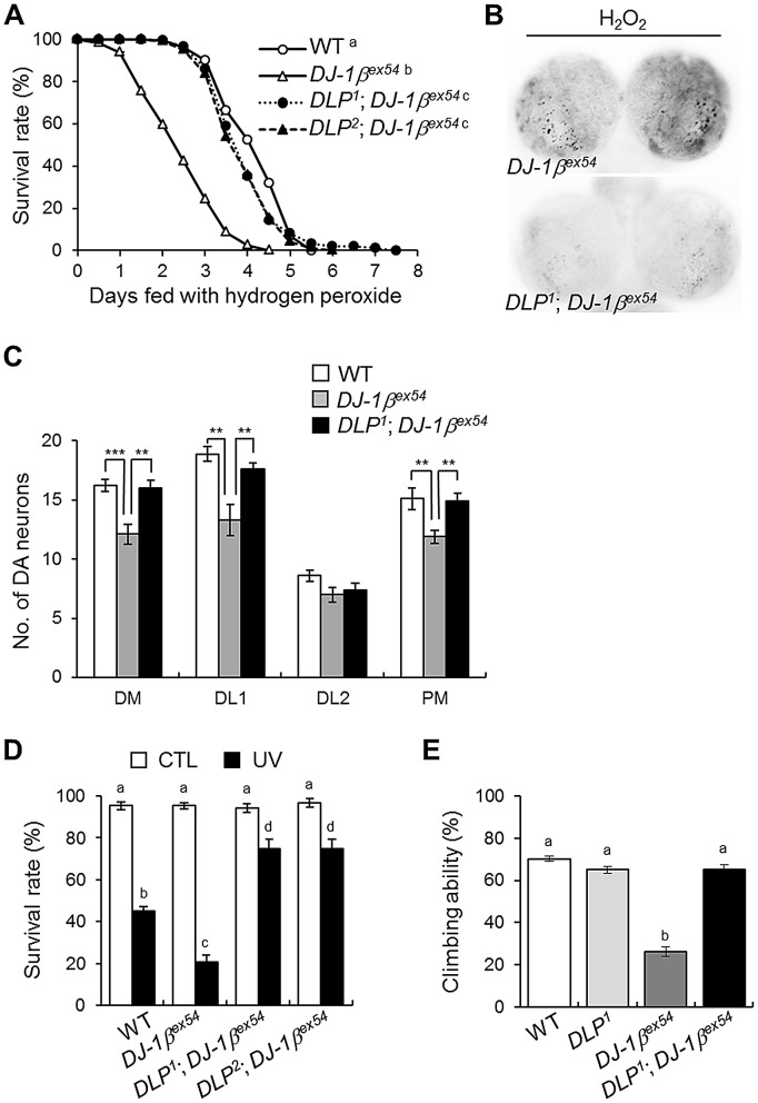
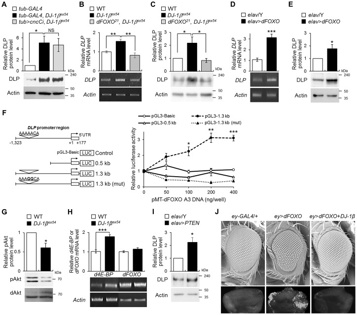
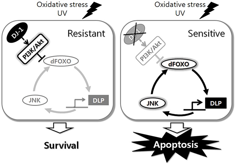
DJ-1, a Parkinson's disease (PD)-associated gene, has been shown to protect against oxidative stress in Drosophila. However, the molecular mechanism underlying oxidative stress-induced phenotypes, including apoptosis, locomotive defects, and lethality, in DJ-1-deficient flies is not fully understood. Here we showed that Daxx-like protein (DLP), a Drosophila homologue of the mammalian Death domain-associated protein (Daxx), was upregulated under oxidative stress conditions in the loss-of-function mutants of Drosophila DJ-1β, a Drosophila homologue of DJ-1. DLP overexpression induced apoptosis via the c-Jun N-terminal kinase (JNK)/Drosophila forkhead box subgroup O (dFOXO) pathway, whereas loss of DLP increased resistance to oxidative stress and UV irradiation. Moreover, the oxidative stress-induced phenotypes of DJ-1β mutants were dramatically rescued by DLP deficiency, suggesting that enhanced expression of DLP contributes to the DJ-1β mutant phenotypes. Interestingly, we found that dFOXO was required for the increase in DLP expression in DJ-1β mutants and that dFOXO activity was increased in the heads of DJ-1β mutants. In addition, subcellular localization of DLP appeared to be influenced by DJ-1 expression so that cytosolic DLP was increased in DJ-1β mutants. Similarly, in mammalian cells, Daxx translocation from the nucleus to the cytosol was suppressed by overexpressed DJ-1β under oxidative stress conditions; and, furthermore, targeted expression of DJ-1β to mitochondria efficiently inhibited the Daxx translocation. Taken together, our findings demonstrate that DJ-1β protects flies against oxidative stress- and UV-induced apoptosis by regulating the subcellular localization and gene expression of DLP, thus implying that Daxx-induced apoptosis is involved in the pathogenesis of DJ-1-associated PD.


Conflict of interest statement

The authors have declared that no competing interests exist.

Figures

Figure 1
Figure 1. Decreased DA neurons and increased apoptosis in DJ-1β mutant under oxidative stress conditions.
(A) DA neurons visualized by immunohistochemical analysis with anti-tyrosine hydroxylase antibody in the brains of wild-type (WT) and DJ-1β mutant (DJ-1βex54) flies fed with 1% H2O2 for 3 days. Dotted boxed areas indicate DA neuron clusters. The lower pictures, including DM, DL1, DL2, and PM, are the magnified 4 dotted boxed areas of the upper pictures. Magnification of the upper pictures, 100×; Magnification of the lower pictures, 400×. (B) Graphs showing the number of DA neurons in each cluster of WT and DJ-1βex54 flies after feeding with H2O2 for 3 days (n = 10, Student's t-test: DM, *** p<0.001; DL1 and PM, ** p<0.01). The data are expressed as means ± s.e. values. (C) Acridine orange staining of 0.1% hydrogen peroxide-treated larval brains showed that increased oxidative stress-induced apoptosis in DJ-1βex54 compared to the WT controls. DM, dorsomedial clusters; DL, dorsolateral clusters; PM, posteriomedial clusters.
Figure 2
Figure 2. Gene expression of DLP by oxidative stress, UV, and DJ-1β.
(A) DLP protein levels in the wild-type (WT) fly body and head (Student's t-test, n = 5, * p<0.05). (B–C) DLP mRNA (B) and protein (C) levels in WT embryos exposed to UV (50 mJ/cm2) and the heads of WT flies fed with 1% H2O2 (B, Student's t-test: UV, n = 5, ** p<0.01; H2O2, n = 5, ** p<0.01; C, Student's t-test: UV, n = 5, * p<0.05; H2O2, n = 4, ** p<0.01). (D–G) DLP mRNA (D, F) and protein (E, G) levels in the heads of WT and DJ-1βex54 flies grown with cornmeal-soybean standard fly food (D–E) and fed with 1% H2O2 for 3 days (F–G). (D, Student's t-test, n = 8; E, Student's t-test, n = 7; F, Student's t-test, n = 6, * p<0.05; G, Student's t-test, n = 7, * p<0.05). (H–I) DLP mRNA (H) and protein (I) levels in control (elav/Y) and pan-neuronally DJ-1β-overexpressing (elav>HA-DJ-1β) fly head (H, Student's t-test, n = 5, *** p<0.001; I, Student's t-test, n = 4, *** p<0.001). (J) Daxx mRNA level in the WT and DJ-1 null SN4741 cells treated with 1 mM H2O2 (Student's t-test, n = 3, ** p<0.01). (K–L) Western blot (K) and statistical (L) analysis of cytosolic (c) and nuclear (n) fractions of WT and DJ-1βex54 fly head extracts showed that increased translocation of DLP to the cytosol in DJ-1βex54 compared to WT controls (Student's t-test, n = 4, ** p<0.01). Relative cytosolic DLP levels were calculated by dividing the normalized cytosolic DLP level by the normalized nuclear DLP level. Lamin and β-tubulin were used as loading controls for nuclear and cytosolic fractions, respectively. (M) Confocal images of DLP immunohistochemistry in the larval brains of WT and DJ-1βex54. Hoechst-stained regions represent nuclei. Magnification, 8,000×. (N) Confocal images showing subcellular localization of Daxx in WT and DJ-1 null SN4741 cells. MitoTracker-stained spots represent mitochondria. The cells were treated with 0.4 mM H2O2 for 1 h. (O) The ratio of the cells with cytosolic localized Daxx in DJ-1 null SN4741 cells transfected with wild-type DJ-1β or nucleus (Nuc)-, cytoplasmic membrane (CM)-, Golgi (Golgi)-, or mitochondria (Mito)-targeted DJ-1β. The cells were treated with 0.4 mM H2O2 for 1 h. More than 70 cells per each samples were counted to calculate the ratio of the cells with cytosolic Daxx (Student's t-test, n = 4, * p<0.05, *** p<0.001). All data are expressed as means ± s.e. values. Actin was employed as an internal control of total extract.
Figure 3
Figure 3. Generation and characterization of DLP mutants.
(A) Genomic structure of the DLP gene. Exons of the DLP gene are shown in black (coding region) and white (non-coding region) boxes. The inverted triangles indicate the P-elements, EY09290 and KG01694. The deletion sites of DLP1, DLP2, DLP3, and DLP4 are illustrated under the genomic structures. (B) Determination of the deleted size in DLP mutants by genomic DNA PCR. (C) Western blotting of DLP in wild type (WT), DLP loss-of-function (DLP1 and DLP2) and gain-of-function (elav>DLP) mutants. Intact DLP protein is not detected in the DLP mutants. (D) Comparison of DLP gene expression in the third-instar larval brains of WT and a DLP mutant via RNA in situ hybridization. (E–F) Survival rates of DLP loss-of-function (DLP1, DLP2, and elav>DLPi) and gain-of-function (elav>DLP) mutants under oxidative stress conditions. (E) WT and DLPrv/DLP1 were used as controls (log-rank test: WT, n = 300; DLP1, n = 250; DLP2, n = 250; DLPrv/DLP1, n = 300, p<0.01, groups with the same letter do not differ significantly). (F) elav/Y was used as a control (log-rank test: elav/Y, n = 350; elav>DLP, n = 300; elav>DLPi, n = 300, p<0.01, groups with the different letter differ significantly). The genotypes of the samples were elav/Y (elav-GAL4/Y), elav>DLP (elav-GAL4/Y; EY09290/+), and elav>DLPi (elav-GAL4/Y; UAS-DLP-RNAi/+). (G) Acridine orange staining of larval brains of DLP1 and WT treated with 0.1% H2O2 for 24 h. (H) Survival rates of WT and DLP mutant (DLP1 and DLP2) pupae after exposure to UV irradiation (10 mJ/cm2; black bars) as described in the Materials and Methods (Kruskal-Wallis test: CTL, n≥6, p<0.1; UV, n = 6, p<0.01, groups with the same letter do not differ significantly). CTL, UV-untreated control pupae; UV, UV-treated pupae. All data are expressed as means ± s.e. values. (I) TUNEL-stained images of UV-exposed 0–3 h embryos of WT and DLP1. The lower panels are higher-magnification images of the boxes indicated with dotted lines in the upper panels. CTL, control; rv, revertant; TUNEL, terminal deoxynucleotidyl transferase-mediated dUTP-biotin nick end-labeling.
Figure 4
Figure 4. DLP activates apoptosis and the JNK/dFOXO signaling pathway.
(A) Comparison of tissue sizes of control (MS1096/Y), DLP-overexpressing (MS1096>DLP and MS1096>DLP ×2), and DLP- and DIAP1-coexpressing (MS1096>DLP ×2+DIAP1) fly wings. Two copies of the DLP gene were overexpressed in MS1096>DLP ×2. (B) Acridine orange-stained images of control (MS1096/Y), DLP-overexpressing (MS1096>DLP and MS1096>DLP ×2), DLP- and hep-coexpressing (MS1096>DLP+hep), DLP-overexpressing and hep deficient (MS1096>DLP, hep1), DLP-overexpressing and bsk deficient (MS1096>DLP, bsk1), DLP- and puc-coexpressing (MS1096>DLP ×2+puc), and DLP-overexpressing and dFOXO deficient (MS1096>DLP ×2, dFOXO21) wing imaginal discs. (C) Genetic interactions of DLP with bsk, hep, and puc in the developing wing. The reduced wing phenotype induced by DLP overexpression (MS1096>DLP) was strongly exacerbated by bsk (MS1096>DLP+bsk) or hep (MS1096>DLP+hep) overexpression, and suppressed by bsk (MS1096>DLP, bsk1) or hep (MS1096>DLP, hep1) deficiency or co-expression of puc (MS1096>DLP ×2+puc). (D) Comparison of JNK activity in the DLP- and bsk-coexpressing or DLP- and hep-coexpressing wing imaginal discs (MS1096>DLP+bsk or MS1096>DLP+hep) with DLP-, bsk- or hep-overexpressing wings (MS1096>bsk or MS1096>hep) by anti-phospho-JNK antibody staining. (E) Genetic interactions of DLP with dFOXO in the developing wing. The wing phenotype of DLP overexpression (MS1096>DLP ×2) was strongly suppressed by dFOXO deficiency (MS1096>DLP ×2, dFOXO21). MS1096 with dFOXO deficiency (MS1096, dFOXO21) was used as controls. (F) DLP protein levels in the control (sev-GAL4) and constitutive active hep-overexpressing (sev>hepCA) fly heads (Student's t-test, n = 9, *** p<0.001). sev, sevenless-GAL4. The data are expressed as means ± s.e. values. The genotypes of the samples were MS1096/Y (MS1096-GAL4/Y), MS1096>bsk (MS1096-GAL4/Y; UAS-bsk/+), MS1096>hep (MS1096-GAL4/Y; UAS-hep/+), MS1096, bsk1 (MS1096-GAL4/Y; bsk1/+), MS1096, hep1 (MS1096-GAL4/hep1), MS1096>DLP (MS1096-GAL4/Y; EY09290/+), MS1096>DLP+bsk (MS1096-GAL4/Y; EY09290/UAS-bsk), MS1096>DLP+hep (MS1096-GAL4/Y; EY09290/UAS-hep), MS1096>DLP, bsk1 (MS1096-GAL4/Y; EY09290/bsk1), MS1096>DLP, hep1 (MS1096-GAL4/hep1; EY09290/+), MS1096>DLP ×2 (MS1096-GAL4/Y; EY09290/EY09290), MS1096>puc (MS1096-GAL4/Y; UAS-puc/+), MS1096>DLP ×2+puc (MS1096-GAL4/Y; EY09290/EY09290; UAS-puc/+), MS1096, dFOXO21 (MS1096-GAL4/Y;; dFOXO21/+), MS1096>DLP ×2, dFOXO21 (MS1096-GAL4/Y; EY09290/EY09290; dFOXO21/+), MS1096>DLP ×2+DIAP1 (MS1096-GAL4/Y; EY09290/EY09290; UAS-DIAP1/+), sev-GAL4 (sev-GAL4/+), and sev>hepCA (sev-GAL4/+; UAS-hepCA/+). bsk, basket; DIAP1, Drosophila inhibitor of apoptosis protein 1; hep, hemipterous; pJNK, phospho-JNK; puc, puckered.
Figure 5
Figure 5. DLP deficiency reduces acute sensitivity to oxidative stress and UV, and improves locomotive dysfunction in DJ-1β mutant.
(A) Comparison of the survival rates of DLP and DJ-1β double mutants (DLP1; DJ-1βex54 and DLP2; DJ-1βex54) with wild-type (WT) and DJ-1βex54 flies under oxidative stress conditions (log-rank test: WT, n = 250; DJ-1βex54, n = 250; DLP1; DJ-1βex54, n = 250; DLP2; DJ-1βex54, n = 250, p<0.01, groups with the same letter do not differ significantly). (B) Reduced oxidative stress-induced apoptosis was noted in the larval brain of the DLP and DJ-1β double mutant (DLP1; DJ-1βex54) compared to DJ-1βex54. The larval brains were treated with 0.1% H2O2 for 24 h and cell death was detected via acridine orange staining. (C) Sensitized DA neuronal death of DJ-1βex54 under oxidative stress conditions was rescued by DLP deficiency. The flies were fed with 1% H2O2 for 3 days. (n = 10, Student's t-test: DM, ** p<0.01, *** p<0.001; DL1, ** p<0.01; PM, ** p<0.01). (D) Survival rates of WT, DJ-1βex54, and double mutant of DLP and DJ-1β (DLP1; DJ-1βex54 and DLP2; DJ-1βex54) pupae after exposure to UV irradiation (10 mJ/cm2; black bars) as described in the Materials and Methods (Kruskal-Wallis test: CTL, n≥6, p>0.1; UV, n≥5, p<0.01, groups with the same letter do not differ significantly). CTL, UV-untreated control pupae; UV, UV-treated pupae. (E) Comparison of climbing abilities of WT, DLP1, DJ-1βex54, and double mutants of DLP and DJ-1β (DLP1; DJ-1βex54). The climbing abilities of 5-day-old flies for each group were tested as described in the Materials and Methods (ANOVA and Tukey's HSD analysis: n≥12, p<0.01, groups with the same letter do not differ significantly). All data are expressed as means ± s.e. values.
Figure 6
Figure 6. The role of dFOXO in the regulation of DLP by DJ-1β.
(A) DLP protein levels in the heads of control (tub-GAL4), DJ-1β mutant (tub-GAL4, DJ-1βex54) and the double mutant of cncC and DJ-1β (tub>cncCi, DJ-1βex54) flies fed with 1% H2O2 (Student's t-test, n = 4, * p<0.05). NS, not significant. (B–C) DLP mRNA (B) and protein (C) levels in the head of WT, DJ-1βex54, and dFOXO and DJ-1β double mutant (dFOXO21, DJ-1βex54) flies fed with 1% H2O2 (B, Student's t-test, n = 5, ** p<0.01; C, Student's t-test, n = 4, * p<0.05). (D–E) DLP mRNA (D) and protein (E) levels in control (elav/Y) and pan-neuronally dFOXO-overexpressing (elav>dFOXO) fly heads (D, Student's t-test, n = 6, *** p<0.001; E, Student's t-test, n = 5, * p<0.05). (F) Luciferase assays showed activation of DLP promoters in S2 cells after cotransfection with dFOXO-A3. (Open circle) Empty vector. (Open triangle) 0.5-kb fragment of DLP promoter. (Filled circle) 1.3-kb fragment of DLP promoter. (Filled triangle) 1.3-kb fragment of DLP promoter with mutation in the putative FRE site: pGL3–1.3 kb (mut). Bold characters in the putative FRE site represent the mutated nucleotides. (Student's t-test, n = 3, * p<0.05; ** p<0.01; *** p<0.001). (G) The levels of phospho-Akt in the head of WT and DJ-1βex54 flies fed with 1% H2O2 (Student's t-test, n = 3, * p<0.05). dAkt was used as an internal control. (H) d4E-BP, a target of dFOXO, and dFOXO mRNA levels in the head of WT and DJ-1βex54 flies fed with 1% H2O2 (Student's t-test: d4E-BP, n = 7, *** p<0.001; dFOXO, n = 7). (I) DLP protein levels in the control (elav/Y) and pan-neuronally PTEN-overexpressing (elav>PTEN) fly heads (Student's t-test, n = 4, * p<0.05). (J) Genetic interactions of dFOXO with DJ-1β in the developing eye. The upper pictures are scanning electron micrographs of the fly eyes. The lower pictures are acridine orange-stained images of the eye imaginal discs. The genotypes of the samples were tub-GAL4 (tub-GAL4/+), tub-GAL4, DJ-1βex54 (tub-GAL4, DJ-1βex54/DJ-1βex54), tub>cncCi, DJ-1βex54 (UAS-cncC-RNAi/+; tub-GAL4, DJ-1βex54/DJ-1βex54), dFOXO21, DJ-1βex54 (dFOXO21, DJ-1βex54/DJ-1βex54), elav/Y (elav-GAL4/Y), elav>dFOXO (elav-GAL4/Y; UAS-dFOXO/+), elav>PTEN (elav-GAL4/Y; UAS-PTEN/+), ey-GAL4 (ey-GAL4/+), ey>dFOXO (UAS-dFOXO/+; ey-GAL4/+), and ey>dFOXO+DJ-1β (UAS-dFOXO/UAS-HA-DJ-1β; ey-GAL4/+). pAkt, phospho-Akt; ey, eyeless. All data are expressed as means ± s.e. values. Actin was used as an internal control.
Figure 7
Figure 7. Schematic representation of the role of Drosophila DJ-1 in the cellular response to oxidative stress or UV.
dFOXO, DLP, and JNK form a circuit that controls cellular responses to stress, and DJ-1 sets neural sensitivity to stress by regulating this circuit.

Similar articles

Cited by

References

    1. Emerit J, Edeas M, Bricaire F (2004) Neurodegenerative diseases and oxidative stress. Biomed Pharmacother 58: 39–46. - PubMed
    1. Lin MT, Beal MF (2006) Mitochondrial dysfunction and oxidative stress in neurodegenerative diseases. Nature 443: 787–795. - PubMed
    1. Tsang AHK, Chung KKK (2009) Oxidative and nitrosative stress in Parkinson's disease. Biochim Biophys Acta 1792: 643–650. - PubMed
    1. Jenner P (2007) Oxidative stress and Parkinson's disease. Handb Clin Neurol 83: 507–520. - PubMed
    1. Zhou C, Huang Y, Przedborski S (2008) Oxidative Stress in Parkinson's Disease. Ann N Y Acad Sci 1147: 93–104. - PMC - PubMed

Publication types

MeSH terms

Substances