. 2013 Apr;9(4):e1003415.
doi: 10.1371/journal.pgen.1003415. Epub 2013 Apr 4.

Intrinsic epigenetic regulation of the D4Z4 macrosatellite repeat in a transgenic mouse model for FSHD

Affiliations

Intrinsic epigenetic regulation of the D4Z4 macrosatellite repeat in a transgenic mouse model for FSHD

Yvonne D Krom et al. PLoS Genet. 2013 Apr.

Abstract

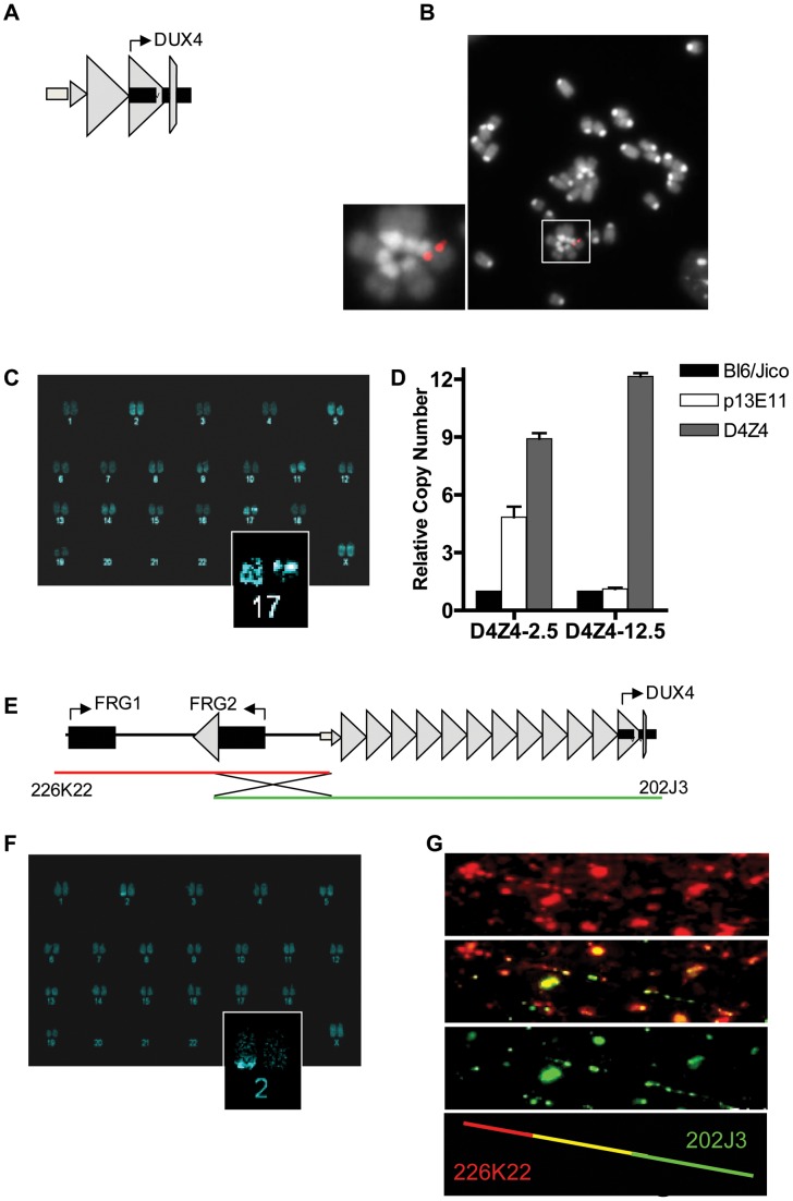
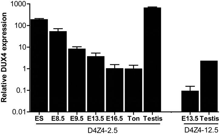
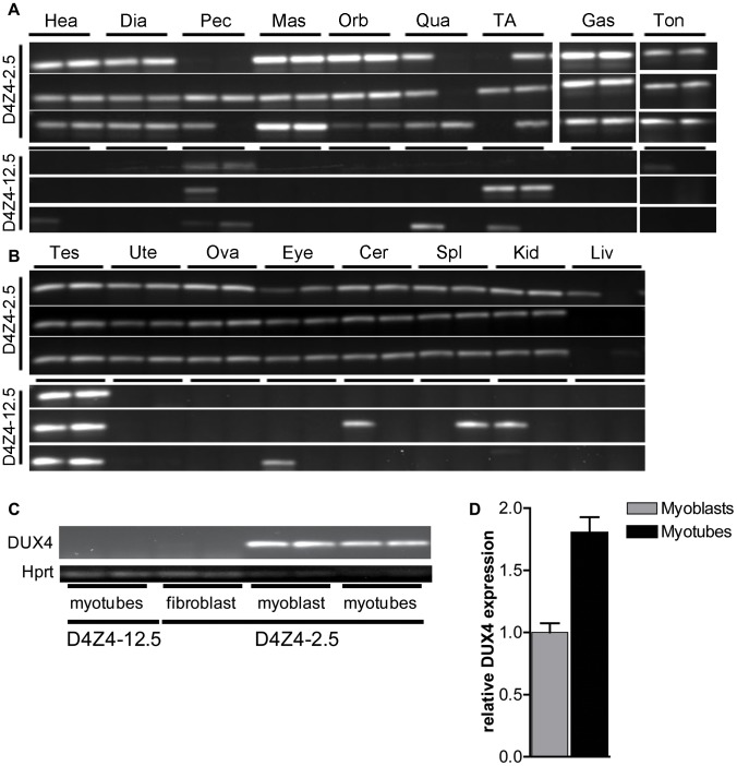
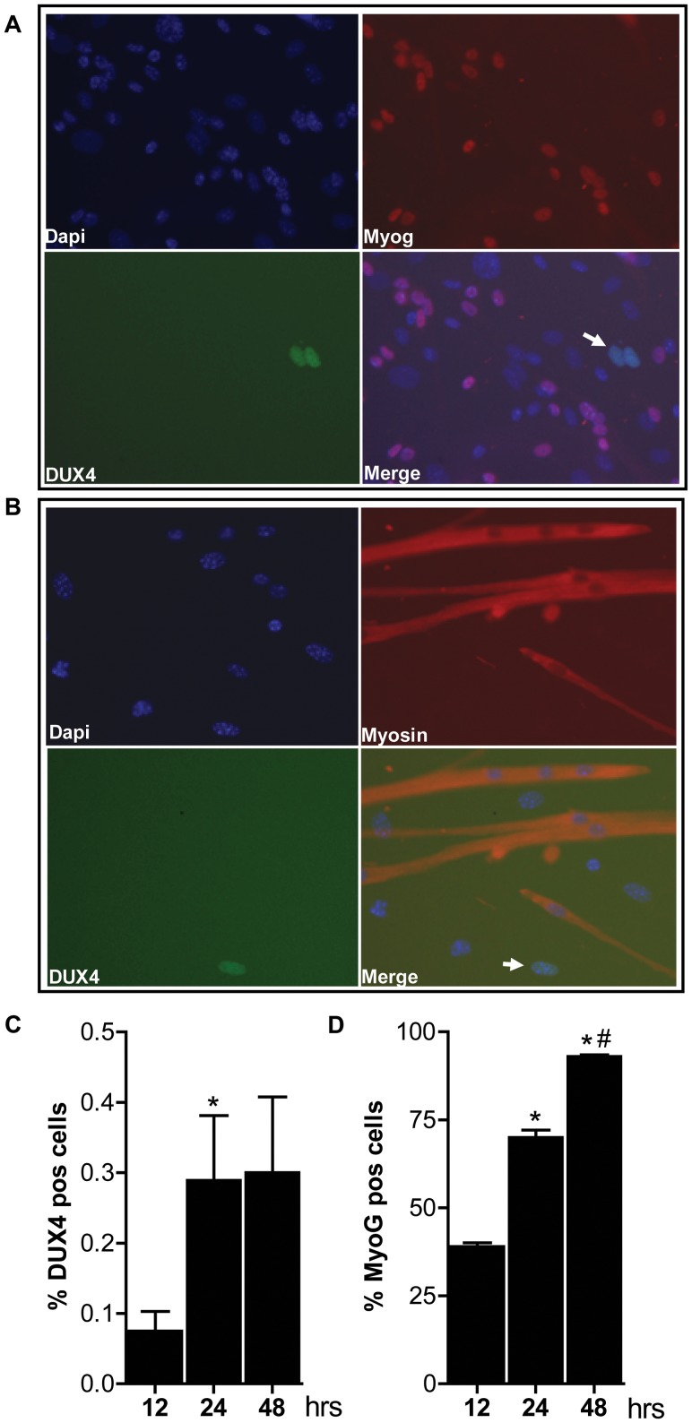
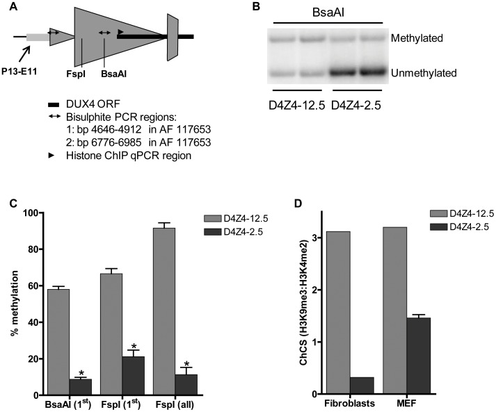
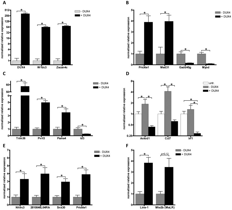
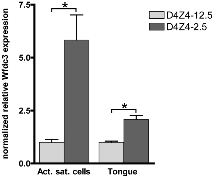
Facioscapulohumeral dystrophy (FSHD) is a progressive muscular dystrophy caused by decreased epigenetic repression of the D4Z4 macrosatellite repeats and ectopic expression of DUX4, a retrogene encoding a germline transcription factor encoded in each repeat. Unaffected individuals generally have more than 10 repeats arrayed in the subtelomeric region of chromosome 4, whereas the most common form of FSHD (FSHD1) is caused by a contraction of the array to fewer than 10 repeats, associated with decreased epigenetic repression and variegated expression of DUX4 in skeletal muscle. We have generated transgenic mice carrying D4Z4 arrays from an FSHD1 allele and from a control allele. These mice recapitulate important epigenetic and DUX4 expression attributes seen in patients and controls, respectively, including high DUX4 expression levels in the germline, (incomplete) epigenetic repression in somatic tissue, and FSHD-specific variegated DUX4 expression in sporadic muscle nuclei associated with D4Z4 chromatin relaxation. In addition we show that DUX4 is able to activate similar functional gene groups in mouse muscle cells as it does in human muscle cells. These transgenic mice therefore represent a valuable animal model for FSHD and will be a useful resource to study the molecular mechanisms underlying FSHD and to test new therapeutic intervention strategies.


Conflict of interest statement

The authors have declared that no competing interests exist.

Figures

Figure 1
Figure 1. Integration site and copy number of D4Z4-2.5 and D4Z4-12.5 constructs in the mouse genome.
A) Schematic draw of the L42 EcoRI fragment used to generate the D4Z4-2.5 mouse line B) Metaphase spread of D4Z4-2.5 fibroblasts co-stained with dapi and the CY3 labeled L42 probe shows integration at a single pair of chromosomes C) COBRA-FISH analysis on D4Z4-2.5 fibroblast metaphase spreads probed with biotinylated-L42 fragments shows integration of L42 on chr17; D) Detection of copy number of the integrated fragments in both mouse models by MLPA analysis. The probe mix contained three probes specific for wild type alleles, one probe designed against the human p13E-11 region and one probe against D4Z4 E) Schematic draw of PAC clones used to generate the D4Z4-12.5 mouse; F) COBRA-FISH analysis on D4Z4-12.5 fibroblast metaphase spreads probed with a biotinylated PAC clone shows integration of the PAC clone on chr2; G) Fiber-FISH analysis of D4Z4-12.5 fibroblasts. Both PAC clones, labeled and hybridized to DNA fibers, were shown to be recombined during integration into the mouse genome.
Figure 2
Figure 2. Quantative expression analysis of DUX4 transcripts from the telomeric D4Z4 unit in D4Z4-12.5 and D4Z4-2.5 mice.
Quantitative RT-PCR data of DUX4 in D4Z4-2.5 embryonic stem cells (ES), in complete embryos of day E8.5, 9.5, 13,5 and 16.5, representing key myogenic developmental stages, and in adult ton = tongue, testis and in complete embryo day 13,5 and testis tissue of D4Z4-12.5 mice. Expression is normalized to the mouse reference gene Hprt and plotted in log10 scale. Error bars indicate SEM of the mean (n = 2–5).
Figure 3
Figure 3. Analysis of transcriptional activity of DUX4 in a panel of tissues of D4Z4-2.5 and D4Z4-12.5 mice.
DUX4 transcripts measured in 7 weeks old D4Z4-2.5 and D4Z4-12.5 mice (n = 3) in A) muscle tissue: Hea = Heart, Dia = Diaphragm, Pec = Pectoralis Mas = Masseter, Orb = Orbicularis oris, Qua = Quadriceps, TA = Tibialis anterior, Gas = Gastrocnemius, Ton = Tongue; and B) somatic non-muscle and germline tissue: Tes = Testis, Ute = Uterus, Ova = Ovarium, Eye, Cer = Cerebellum, Spl = Spleen, Kid = Kidney, Liv = Liver C) DUX4 transcripts measured in satellite-cell-derived myoblasts, myotubes and interstitial fibroblast extracted from EDL muscle of D4Z4-12.5 and D4Z4-2.5 transgenic mice. D) Quantitative RT-PCR data of DUX4 expression in D4Z4-2.5 myoblasts (n = 2) and myotubes (n = 2) 48 hours after induction of differentiation. Errors indicate SEM of the plotted mean.
Figure 4
Figure 4. Bursts of DUX4 protein expression in differentiating D4Z4-2.5 muscle cells.
Satellite-cell-derived myoblasts extracted from single EDL fibers of D4Z4-2.5 mice were differentiated for 12, 24 and 48 hrs and co-stained for DUX4 and Myog or for DUX4 and Myosin heavy chain. A) Representative DUX4 and Myog IF staining images of D4Z4-2.5 myotubes, 24 hrs after induction of differentiation, indicate absence of Myog in DUX4 expressing cells. B) Representative DUX4 and Myosin HC IF staining images of D4Z4-2.5 myotubes, 24 hrs after induction of differentiation, indicate exclusion of DUX4 positive cells from newly formed myotubes. Both DUX4 (panel C) and Myog (panel D) positive nuclei in relation to total amount of nuclei (DAPI staining) were counted during the differentiation process. C) Approximately 2∶1000 nuclei showed nuclear DUX4 staining. D) The percentage of Myog positive nuclei revealed an increase in differentiation committed cells with time. After 48 hours of differentiation almost all myoblasts are committed to differentiation. Error bars indicate stdev of the plotted mean (n = 7); *p<0,05 compared to t = 12 hrs; #p<0,05 compared to t = 24 hrs.
Figure 5
Figure 5. Epigenetic structure of D4Z4 in D4Z4-2.5 and D4Z4-12.5 mice.
A) Schematic draw of the regions within D4Z4 where CpG and histone methylation were interrogated. B) Representative figure of a methylation sensitive Southern blot assay to quantify DNA methylation levels. Upon BsaAI digestion, gel separation and blotting, two distinct bands representing the unmethylated and methylated fragment are visualized and quantified; C) Southern Blot analysis was done using two different methylation sensitive restriction enzymes, BsaAI and FspI, in adult gastrocnemius muscle tissue of D4Z4-12.5 and D4Z4-2.5 mice. Both probes p13E-11 and D4Z4 were used to measure CpG methylation levels in the most proximal unit and all units, respectively. The methylation percentages of the two different CpG sites are plotted. Error bars indicate stdev of the plotted mean (n = 4 D4Z4-12.5 vs n = 5 D4Z4-2.5, *p<0.001). D) Histone methylation levels of D4Z4 in transgenic D4Z4-12.5 and D4Z4-2.5 embryonic (MEFs) and adult fibroblasts. Chromatin was precipitated with H3K4me2, H3K9me3 and control IgG antibodies. Precipitated DNA was amplified with qPCR primers amplifying the transcription start site of DUX4. Levels of H3K9me3 in relation to H3K4me2 have been plotted as the chromatin compaction score (ChCS).
Figure 6
Figure 6. Validation of expression levels of DUX4 deregulated genes in C2C12 myoblasts.
A set of deregulated genes obtained from expression array analysis was confirmed by qRT-PCR. Expression analysis of A) DUX4 and genes that are switched on by DUX4 in C2C12 cells, B) genes that respond to DUX4 in humans and mice, C) germ line and early development associated genes, D) innate immunity genes, untr = untransfected control, transfection activates innate immunity which is dampened by DUX4 expression, E) genes directly regulated by DUX4 which were identified by ChIP-seq and F) activated L1 and MaLR retrotransposons. For panel A and Mte2b in panel F, DUX4- values refer to the DUX4 depleted FACS sorted fraction, enabling proper normalization of genes switched on upon DUX4 expression. In all other panels DUX4- refers to pCS2 transfected cells. All expression levels are relative to Cyclophillin-B and normalized to DUX4- or wt conditions. Error bars indicate SEM of at least triplicate measurements, asterisks indicate p-values<0.05 based on a student's t-test (panels A, B, C, E & F) or one way ANOVA (panel D) analysis.
Figure 7
Figure 7. Expression of the DUX4 induced Wfdc3 gene in myoblasts and tongue muscle of D4Z4-2.5 mice.
Relative expression of Wfdc3 in satellite-cell-derived myoblast cultures from single EDL fibers (D4Z4-2.5: n = 6 and D4Z4-12.5: n = 5) and tongue tissue isolated from 7–8 weeks old mice (D4Z4-2.5: n = 6 and D4Z4-12.5: n = 6). Expression levels are relative to Cyclophilin-B and Hprt and normalized to levels in D4Z4-12.5 mice, plotted as the mean ± SEM. Asterisks indicate p<0.05 according to a independent two-tailed student's t-test.

Similar articles

Cited by

References

    1. Clapp J, Mitchell LM, Bolland DJ, Fantes J, Corcoran AE, et al. (2007) Evolutionary conservation of a coding function for D4Z4, the tandem DNA repeat mutated in facioscapulohumeral muscular dystrophy. Am J Hum Genet 81: 264–279 S0002-9297(07)61193-8 [pii];10.1086/519311 [doi]. - PMC - PubMed
    1. Dixit M, Ansseau E, Tassin A, Winokur S, Shi R, et al. (2007) DUX4, a candidate gene of facioscapulohumeral muscular dystrophy, encodes a transcriptional activator of PITX1. Proc Natl Acad Sci U S A 104: 18157–18162. - PMC - PubMed
    1. Gabriels J, Beckers MC, Ding H, De Vriese A, Plaisance S, et al. (1999) Nucleotide sequence of the partially deleted D4Z4 locus in a patient with FSHD identifies a putative gene within each 3.3 kb element. Gene 236: 25–32. - PubMed
    1. Lyle R, Wright TJ, Clark LN, Hewitt JE (1995) The FSHD-associated repeat, D4Z4, is a member of a dispersed family of homeobox-containing repeats, subsets of which are clustered on the short arms of the acrocentric chromosomes. Genomics 28: 389–397. - PubMed
    1. Geng LN, Yao Z, Snider L, Fong AP, Cech JN, et al. (2012) DUX4 activates germline genes, retroelements, and immune mediators: implications for facioscapulohumeral dystrophy. Dev Cell 22: 38–51 S1534-5807(11)00523-5 [pii];10.1016/j.devcel.2011.11.013 [doi]. - PMC - PubMed

Publication types

MeSH terms