. 2013 Apr;9(4):e1003428.
doi: 10.1371/journal.pgen.1003428. Epub 2013 Apr 4.

An essential role for zygotic expression in the pre-cellular Drosophila embryo

Affiliations

An essential role for zygotic expression in the pre-cellular Drosophila embryo

Zehra Ali-Murthy et al. PLoS Genet. 2013 Apr.

Abstract

The Drosophila embryo proceeds through thirteen mitotic divisions as a syncytium. Its nuclei distribute in the embryo's interior during the first six divisions, dividing synchronously with a cycle time of less than ten minutes. After seven divisions (nuclear cycle 8), the syncytial blastoderm forms as the nuclei approach the embryo surface and slow their cycle time; subsequent divisions proceed in waves that initiate at the poles. Because genetic studies have not identified zygotic mutants that affect the early divisions and because transcription has not been detected before cycle 8, the early, pre-blastoderm embryo has been considered to rely entirely on maternal contributions and to be transcriptionally silent. Our studies identified several abnormal phenotypes in live engrailed (en) mutant embryos prior to cycle 8, as well as a small group of genes that are transcribed in embryos prior to cycle 7. Nuclei in en embryos divide asynchronously, an abnormality that was detected as early as nuclear cycle 2-3. Anti-En antibody detected nuclear En protein in embryos at cycle 2, and expression of an En:GFP fusion protein encoded in the paternal genome was also detected in cycle 2 nuclei. These findings demonstrate that the Drosophila embryo is functionally competent for gene expression prior to the onset of its rapid nuclear divisions and that the embryo requires functions that are expressed in the zygote in order to faithfully prosecute its early, pre-cellularization mitotic cycles.


Conflict of interest statement

MBE is a cofounder and member of the Board of Directors of PLOS. The other authors have declared that no competing interests exist.

Figures

Figure 1
Figure 1. engrailed mutant phenotypes in pre-cellular embryos.
(A) Table: Numbers of nuclear cycle 10 embryos from an enLA4/CyO×enLA7/CyO cross that were selected according to the position of their pole cells relative to the dorsal midline; most that extruded pole cells at the midline (center) developed as en+; most with pole cells offset from the midline developed as en mutants. The buds that appeared at cycle 8 (photos of live embryos, DIC optics) were centered in en+ embryos and offset in en mutants (black arrows indicate buds; white arrows indicate areas of “cytoplasmic clearing”). (B) Fixed cycle 7 embryos from an enLA4/CyO×enLA7/CyO cross, stained with DAPI and anti-En antibody. All nuclei in the normal embryo (left) stain with anti-En antibody and are in prophase (inset a higher magnification view of the two nuclei in area demarcated by dashed white lines); nuclei in the en mutant embryo (right) did not stain with anti-En antibody and are either in prophase (e.g., lower nucleus in area inside dashed white lines) or pre-metaphase (e.g., upper nucleus). Arrows indicate areas of “cytoplasmic clearing” that are approximately centered in the normal embryo and offset in the mutant. (C) Two live embryos endowed with His-RFP imaged with epi-fluorescent optics at cycle 8 (upper panels) and approximately ten minutes later (lower panels). Orientation: anterior, left and dorsal, up. After viewing, both embryos were incubated to 24 hrs after egg laying for genotyping; embryo on left developed normally; embryo on right was en. Nuclei in the normal embryo are synchronous (cycle 8: late interphase/prophase; cycle 9: prophase) and evenly distributed; nuclei in the en mutant are neither synchronous (cycle 8: metaphase/anaphase (m/a), prophase (p); cycle 9: metaphase/anaphase, prophase) nor evenly distributed. White dashed lines approximate boundaries between regions at different cell cycle phases.
Figure 2
Figure 2. Abnormal mitotic nuclei in cycle 2–4 engrailed mutant embryos.
Progeny from a cross of Df(en)/+ parentals were stained with antibodies directed against nuclear Lamin and phospho-Histone 3 (pH3). Nuclei in the normals are synchronous (cycle 2: prophase; cycle 3: pro-metaphase; cycle 4: prophase/metaphase); nuclei in the en mutants are not synchronous (cycle 2–3: two prophase, one anaphase (arrow); cycle 3–4: four late interphase, two metaphase/anaphase (arrows); cycle 4: one prophase/metaphase (arrow), three prophase, two late interphase (arrowheads), two telophase/early interphase (abnormal staining and morphology, star).
Figure 3
Figure 3. Engrailed protein is present in nuclei of pre-blastoderm embryos.
Progeny from a cross of Df(en)/+ parentals were stained with antibodies directed against phospho-Histone 3 (pH3) and Engrailed (En). Nuclei in the normals stain with anti-En and are synchronous (cycle 2, prophase; cycle 3, prophase; cycle 4, prophase/metaphase; cycle 5, prophase/metaphase (two nuclei almost superimposed in this projection image)). Nuclei in the en mutants did not stain with anti-En and are not synchronous (cycle 2–3: one, metaphase (arrowhead), two telophase; cycle 3–4: one, anaphase/telophase (arrowhead), three metaphase; cycle 4: three, interphase (arrowheads), five metaphase; cycle 5–6: eight metaphase (arrowheads), eight telophase).
Figure 4
Figure 4. Engrailed protein is expressed by the paternal genome in pre-blastoderm embryos.
Progeny from a cross of wildtype females with males carrying a 21 kb genomic BAC that had been recombineered to express an En:GFP fusion protein regulated by the en promoter region. Embryos were stained with DAPI and anti-GFP antibody; antigen in the nuclei of approximately half the embryos was detected by staining with anti-GFP antibody. Left two columns: GFP-negative embryos cycles 2 (prophase), 3 (prophase) and 4 (prophase/metaphase); right two columns: GFP-positive, En-expressing embryos cycles 2, 3 and 4 (all prophase).
Figure 5
Figure 5. Q–PCR analysis detects transcription in pre-blastoderm embryos.
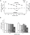
(A) RNA extracted from wildtype embryos at the indicated cycles, from unfertilized eggs (0) and from Df(en) cycle 14 – germ band extended embryos (GBE) was used to template PCR using primers that nestle en intron 1 (see Table S1). No product was detected after 40 PCR cycles using RNA extracted from unfertilized eggs or from Df(en) cycle 10–12 embryos. Product using primers for actin transcripts was detected in all samples. (B) cDNAs prepared from RNA extracted from wildtype embryos at the indicated nuclear cycles was used to template PCR using primers that nestle an intron of en, h, odd, Kr, and eve. Amount of RNA used to prepare the cDNA templates for PCR amplification: (A) 30 ng for nuclear cycles 0–9; 5 ng for nuclear cycles 9–14 and GBE; (B) 30 ng for nuclear cycles 3–6 and 8. PCR reactions were performed in an ABI 3730 Thermocycler for (A) and a BioRad C1000 Touch Thermal Cycler for (B); the number of PCR cycles required to amplify en transcripts differed with the two instruments (presumably due to distinct cycling protocols), but was reproducible for each instrument.
Figure 6
Figure 6. Both homologs express engrailed transcripts in pre-blastoderm embryos.
RNA extracted from embryos heterozygous for a SNP at en transcript nucleotide 1571 (parental sequences shown) was amplified by PCR using primers that nestle en intron 1 (indicted by arrows below drawing of en transcript). Electrophoretic mobility of the PCR product (picture, lower left) was consistent with predicted length (336 bp) for a spliced mRNA template, and the sequence trace (lower right) indicates approximately equal amounts of both parental sequences at the 1571 SNP.
Figure 7
Figure 7. Phenotype of even-skipped pre-blastoderm embryos.
Photographs taken at the indicated intervals of two embryos from a cross of Df(2R)eve/+ parents. (left panels), an embryo with normal pole cell bud formation and with no bulges at the anterior pole prior to cycle 9–10. (top), telophase cycle 8; (bottom), eleven minutes later, cycle 9–10 (right panels), an embryo with bulges at the anterior pole (top) that preceded by eleven minutes the appearance of pole buds. Left embryo developed wildtype denticle belts; right embryo developed denticle belts with the eve “lawn” phenotype.

References

    1. Pritchard DK, Schubiger G (1996) Activation of transcription in Drosophila embryos is a gradual process mediated by the nucleocytoplasmic ratio. Genes Dev 10: 1131–1142. - PubMed
    1. ten Bosch JR, Benavides JA, Cline TW (2006) The TAGteam DNA motif controls the timing of Drosophila pre-blastoderm transcription. Development 133: 1967–1977. - PubMed
    1. Anderson KV, Lengyel JA (1981) Changing rates of DNA and RNA synthesis in Drosophila embryos. Dev Biol 82: 127–138. - PubMed
    1. Edgar BA, Schubiger G (1986) Parameters controlling transcriptional activation during early Drosophila development. Cell 44: 871–877. - PubMed
    1. Zalokar M (1976) Autoradiographic study of protein and RNA formation during early development of Drosophila eggs. Dev Biol 49: 425–437. - PubMed

Publication types

MeSH terms