. 2013 Apr 8;8(4):e59516.
doi: 10.1371/journal.pone.0059516. Print 2013.

Protein trans-splicing of multiple atypical split inteins engineered from natural inteins

Affiliations

Protein trans-splicing of multiple atypical split inteins engineered from natural inteins

Ying Lin et al. PLoS One. .

Abstract

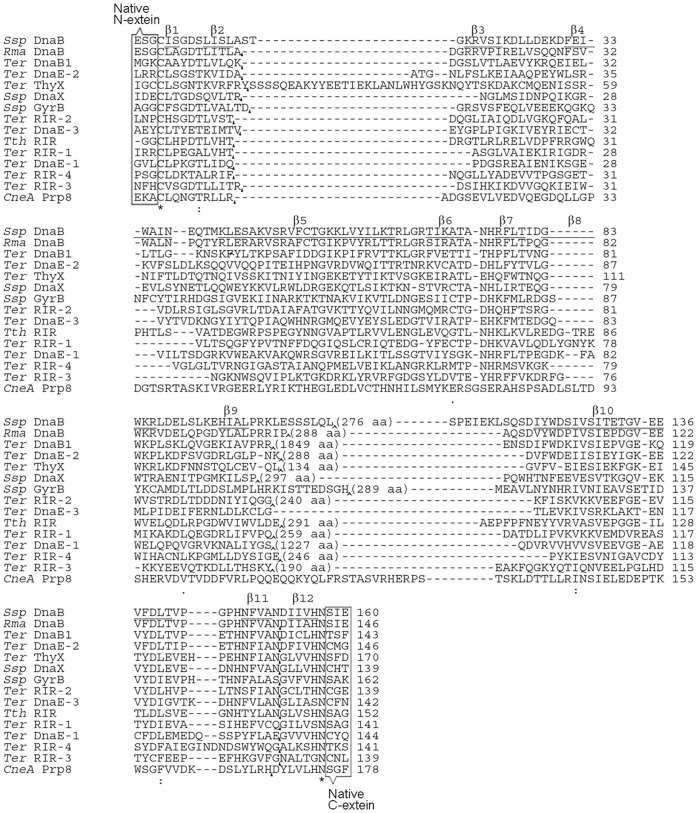
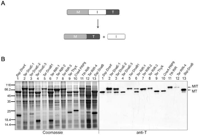
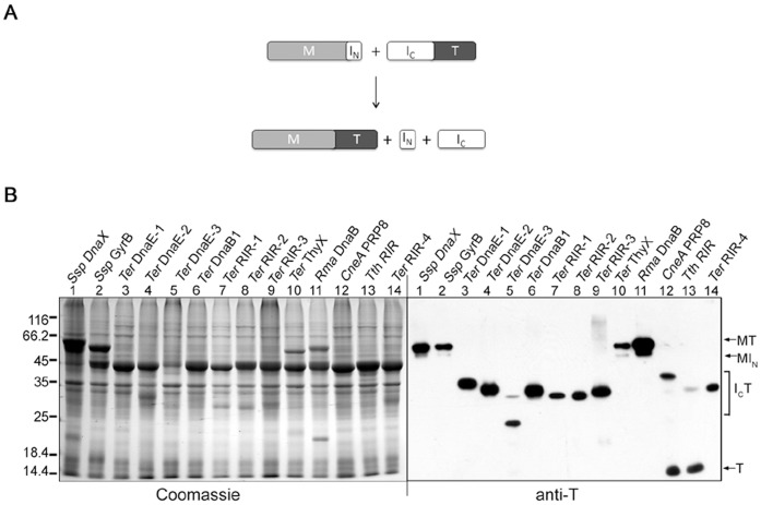
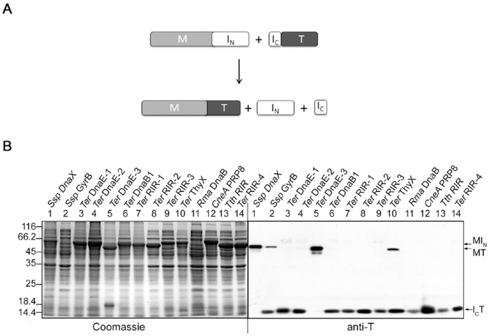
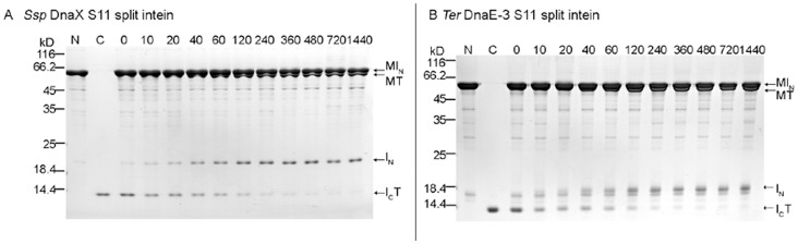
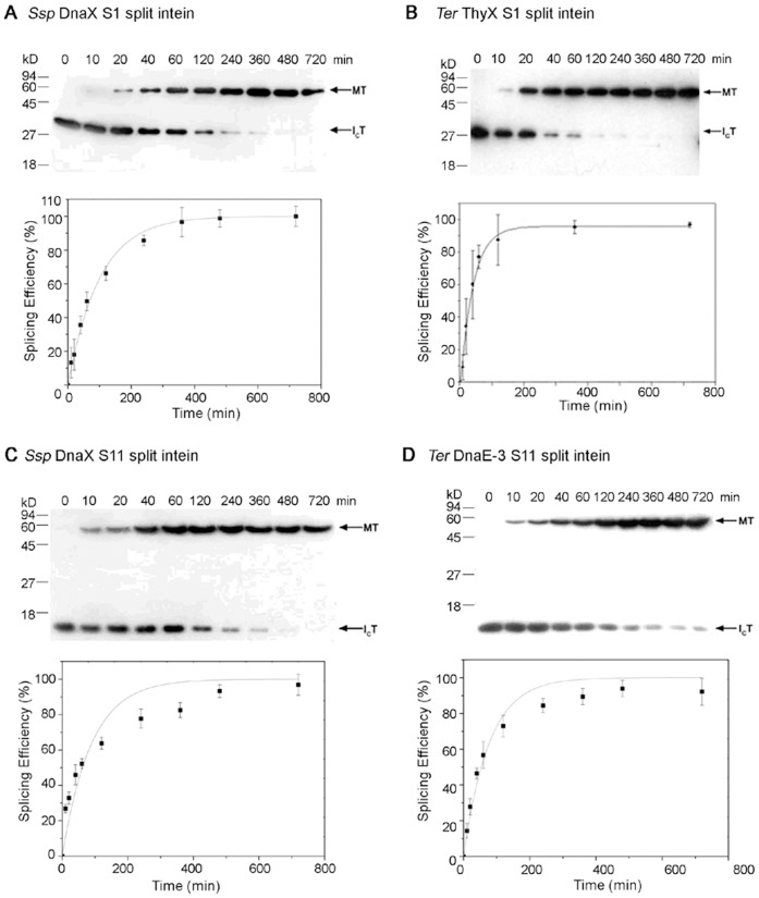
Protein trans-splicing by split inteins has many uses in protein production and research. Splicing proteins with synthetic peptides, which employs atypical split inteins, is particularly useful for site-specific protein modifications and labeling, because the synthetic peptide can be made to contain a variety of unnatural amino acids and chemical modifications. For this purpose, atypical split inteins need to be engineered to have a small N-intein or C-intein fragment that can be more easily included in a synthetic peptide that also contains a small extein to be trans-spliced onto target proteins. Here we have successfully engineered multiple atypical split inteins capable of protein trans-splicing, by modifying and testing more than a dozen natural inteins. These included both S1 split inteins having a very small (11-12 aa) N-intein fragment and S11 split inteins having a very small (6 aa) C-intein fragment. Four of the new S1 and S11 split inteins showed high efficiencies (85-100%) of protein trans-splicing both in E. coli cells and in vitro. Under in vitro conditions, they exhibited reaction rate constants ranging from ~1.7 × 10(-4) s(-1) to ~3.8 × 10(-4) s(-1), which are comparable to or higher than those of previously reported atypical split inteins. These findings should facilitate a more general use of trans-splicing between proteins and synthetic peptides, by expanding the availability of different atypical split inteins. They also have implications on understanding the structure-function relationship of atypical split inteins, particularly in terms of intein fragment complementation.


Conflict of interest statement

Competing Interests: The authors have declared that no competing interests exist.

Figures

Figure 1
Figure 1. Amino acid sequences of mini-inteins and split inteins.
Mini-intein sequences are aligned using ClustalW online , and gaps (represented by -) were introduced to optimize the alignment. CneA PRP8 intein was a natural mini-intein. Ter DnaE-3 mini-intein was derived from a natural conventional split intein by a fusion of the intein fragments. Other mini-inteins were derived from natural inteins by a deletion of their putative endonuclease domain sequences, with the position and number of deleted residues shown in parenthesis. A linker sequence (ASGHHHHHHGGSGS) was inserted at the site of deletion (or corresponding site in the CneA PRP8 and the Ter DnaE-3 mini-inteins) and marked with an arrowhead. For each intein, three (or two) amino acid residues (enclosed with a rectangle) of the native extein sequences on each side of the intein were included in all splicing studies. In the Ssp DnaB mini-intein, whose crystal structure is known, sequences of the 12 β-strands (β1 to β12) are underlined. Split sites for producing the S1 and S11 split inteins are marked with black triangles.
Figure 2
Figure 2. Mini-intein cis-splicing in E. coli cells.
A. Schematic illustration of the cis-splicing reaction. The recombinant precursor protein consists of a maltose binding protein sequence (M) followed by the mini-intein sequence (I) and a thioredoxin sequence (T). B. Detection of splicing activity. After expression of the precursor protein containing a mini-intein (specified on top) in E. coli for overnight at 25°C, total cellular proteins were resolved by SDS-PAGE, followed by Western blotting using an anti-thioredoxin (anti-T) antibody that detected the precursor protein (MIT) and the spliced protein (MT).
Figure 3
Figure 3. S1 split intein trans-splicing in E. coli cells.
A. Schematic illustration of the trans-splicing reaction. Recombinant N-protein consists of a maltose binding protein sequence (M) followed by the small N-intein (IN). Recombinant C-protein consists of the larger C-intein (IC) followed by a thioredoxin (T). B. Detection of trans-splicing. For each S1 split intein (specified on top), the N-protein and the C-protein were co-expressed in E. coli for overnight at 25°C, total cellular proteins were resolved by SDS-PAGE, and protein bands were visualized by Coomassie Blue staining or by Western blotting using an anti-T antibody as indicated. Positions are marked for the spliced protein (MT), the N-protein (MIN), the C-protein (ICT), and a C-cleavage product (T).
Figure 4
Figure 4. S11 split intein trans-splicing in E. coli cells.
A. Schematic illustration of the trans-splicing reaction. Recombinant N-protein consists of a maltose binding protein sequence (M) followed by the large N-intein (IN). Recombinant C-protein consists of the small C-intein (IC) followed by a thioredoxin (T). B. Detection of trans-splicing. For each S11 split intein (specified on top), the N-protein and the C-protein were co-expressed in E. coli for overnight at 25°C, total cellular proteins were resolved by SDS-PAGE, and protein bands were visualized by Coomassie Blue staining or by Western blotting using an anti-T antibody as indicated. Positions are marked for the spliced protein (MT), the N-protein (MIN), and the C-protein (ICT).
Figure 5
Figure 5. S11 split intein trans-splicing in vitro.
For each split intein, purified C-protein (lane C) and N-protein (lane N), which are illustrated in Figure 4A, were mixed in a 1∶5 molar ratio and incubated under fixed set of conditions (25°C, 250 mM NaCl, 1 mM DTT, 20 mM Tris-HCl, pH 8.0). After specified number of minutes (specified on top), samples were taken and analyzed by SDS-PAGE and visualized by Coomassie blue staining. Positions are indicated for the precursor proteins (MIN and ICT), the splicing product (MT), and the excised N-intein (IN). The excised C-intein was too small (6 aa) to be seen in this analysis. Size markers (kD) are shown on the left.
Figure 6
Figure 6. Kinetic analysis of trans-splicing in vitro.
For each S1 or S11 split intein (specified in A to D), purified C-protein and N-protein (illustrated in Figures 3A and 4A) were mixed in a 1∶10 molar ratio and incubated under same conditions as in Figure 5. Samples were taken at different times (specified on top) and analyzed by Western blotting using an anti-T antibody. From the Western blot, relative amounts (band density) of the spliced protein (MT) and the C-protein (ICT) were estimated, and the splicing efficiency was calculated as MT/(MT+ICT). The splicing efficiency was plotted against the reaction time, which was used to estimate the reaction rate constant. All experiments were performed in triplicate, and error bars represent standard deviation.

Similar articles

Cited by

References

    1. Perler FB, Davis EO, Dean GE, Gimble FS, Jack WE, et al. (1994) Protein splicing elements: inteins and exteins–a definition of terms and recommended nomenclature. Nucleic Acids Res 22: 1125–1127. - PMC - PubMed
    1. Perler FB (2002) InBase: the Intein Database. Nucleic Acids Res 30: 383–384. - PMC - PubMed
    1. Chong S, Shao Y, Paulus H, Benner J, Perler FB, et al. (1996) Protein splicing involving the Saccharomyces cerevisiae VMA intein. The steps in the splicing pathway, side reactions leading to protein cleavage, and establishment of an in vitro splicing system. J Biol Chem 271: 22159–22168. - PubMed
    1. Liu XQ (2000) Protein-splicing intein: Genetic mobility, origin, and evolution. Annu Rev Genet 34: 61–76. - PubMed
    1. Hall TM, Porter JA, Young KE, Koonin EV, Beachy PA, et al. (1997) Crystal structure of a Hedgehog autoprocessing domain: homology between Hedgehog and self-splicing proteins. Cell 91: 85–97. - PubMed

Publication types