

Short-latency afferent inhibition modulation during finger movement
- PMID: 23593228
- PMCID: PMC3617156
- DOI: 10.1371/journal.pone.0060496
Short-latency afferent inhibition modulation during finger movement
Abstract
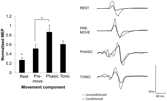
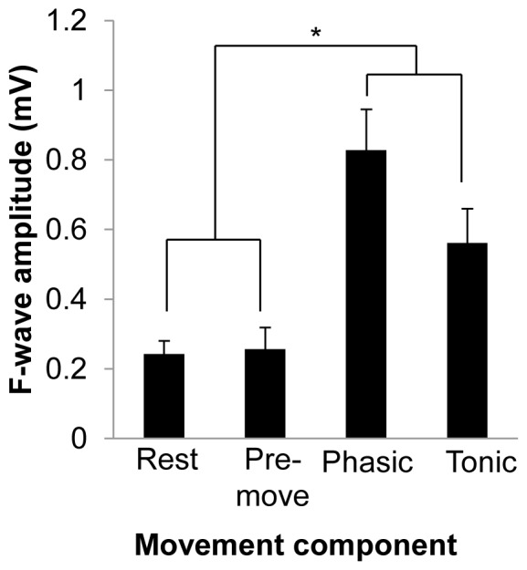
When somatosensory input via electrical stimulation of a peripheral nerve precedes a transcranial magnetic stimulation (TMS) pulse over the primary motor cortex (M1) the corticospinal output is substantially reduced, a phenomenon known as short-latency afferent inhibition (SAI). The present study investigated SAI during rest and during pre-movement, phasic and tonic components of movement. Participants were required to perform an index finger flexion reaction time task in response to an auditory cue. In a series of experiments, SAI was evoked from the mixed, median nerve at the wrist or the cutaneous, digital nerve stimulation of the index finger. To assess the spinal versus cortical origin of movement-related modulation of SAI, F-wave amplitudes were measured during rest and the three movement components. Results indicated that SAI was reduced during all movement components compared to rest, an effect that occurred for both nerves stimulated. Pre-movement SAI reduction was primarily attributed to reduced cortical inhibition, while increased spinal excitability additionally contributed to reduced SAI during tonic and phasic components of movement. SAI was differentially modulated across movement components with mixed but not cutaneous nerve stimulation. These findings reveal that SAI is reduced during movement and this reduction begins as early as the preparation to move. Further, these data suggest that the degree of SAI reduction during movement may be specific to the volume and/or composition of afferent input carried by each nerve.
Conflict of interest statement
Figures


Similar articles
-
Short-latency afferent inhibition determined by the sensory afferent volley.J Neurophysiol. 2016 Aug 1;116(2):637-44. doi: 10.1152/jn.00276.2016. Epub 2016 May 25. J Neurophysiol. 2016. PMID: 27226451 Free PMC article.
-
Biological sex differences in afferent-mediated inhibition of motor responses evoked by TMS.Brain Res. 2021 Nov 15;1771:147657. doi: 10.1016/j.brainres.2021.147657. Epub 2021 Sep 9. Brain Res. 2021. PMID: 34509460
-
Modulation of short-latency afferent inhibition depends on digit and task-relevance.PLoS One. 2014 Aug 13;9(8):e104807. doi: 10.1371/journal.pone.0104807. eCollection 2014. PLoS One. 2014. PMID: 25118700 Free PMC article.
-
Association of short- and long-latency afferent inhibition with human behavior.Clin Neurophysiol. 2021 Jul;132(7):1462-1480. doi: 10.1016/j.clinph.2021.02.402. Epub 2021 Apr 21. Clin Neurophysiol. 2021. PMID: 34030051 Review.
-
Short- and long-latency afferent inhibition; uses, mechanisms and influencing factors.Brain Stimul. 2018 Jan-Feb;11(1):59-74. doi: 10.1016/j.brs.2017.09.009. Epub 2017 Sep 20. Brain Stimul. 2018. PMID: 28964754 Review.
Cited by
-
Preserved central cholinergic functioning to transcranial magnetic stimulation in de novo patients with celiac disease.PLoS One. 2021 Dec 16;16(12):e0261373. doi: 10.1371/journal.pone.0261373. eCollection 2021. PLoS One. 2021. PMID: 34914787 Free PMC article.
-
Short-latency afferent inhibition in chronic spinal cord injury.Transl Neurosci. 2015 Nov 26;6(1):235-243. doi: 10.1515/tnsci-2015-0025. eCollection 2015. Transl Neurosci. 2015. PMID: 28123808 Free PMC article.
-
Clinical diagnostic utility of transcranial magnetic stimulation in neurological disorders. Updated report of an IFCN committee.Clin Neurophysiol. 2023 Jun;150:131-175. doi: 10.1016/j.clinph.2023.03.010. Epub 2023 Mar 29. Clin Neurophysiol. 2023. PMID: 37068329 Free PMC article. Review.
-
Short-latency afferent inhibition determined by the sensory afferent volley.J Neurophysiol. 2016 Aug 1;116(2):637-44. doi: 10.1152/jn.00276.2016. Epub 2016 May 25. J Neurophysiol. 2016. PMID: 27226451 Free PMC article.
-
Experimental environment improves the reliability of short-latency afferent inhibition.PLoS One. 2023 Feb 22;18(2):e0281867. doi: 10.1371/journal.pone.0281867. eCollection 2023. PLoS One. 2023. PMID: 36812217 Free PMC article.
References
-
- Iriki A, Pavlides C, Keller A, Asanuma H (1989) Long-term potentiation in the motor cortex. Science 245: 1385–1387. - PubMed
-
- Swadlow HA (1994) Efferent neurons and suspected interneurons in motor cortex of the awake rabbit: axonal properties, sensory receptive fields, and subthreshold synaptic inputs. J Neurophysiol 71: 437–453. - PubMed
-
- Padel Y, Relova JL (1991) Somatosensory responses in the cat motor cortex. I. Identification and course of an afferent pathway. J Neurophysiol 66: 2041–2058. - PubMed
-
- Jones EG, Powell TP (1969) The cortical projection of the ventroposterior nucleus of the thalamus in the cat. Brain Res 13: 298–318. - PubMed
Publication types
MeSH terms
LinkOut - more resources
Full Text Sources
Other Literature Sources
