. 2013 Apr 8;8(4):e60518.
doi: 10.1371/journal.pone.0060518. Print 2013.

Fiber pathway pathology, synapse loss and decline of cortical function in schizophrenia

Affiliations

Fiber pathway pathology, synapse loss and decline of cortical function in schizophrenia

Max R Bennett et al. PLoS One. .

Abstract

A quantitative cortical model is developed, based on both computational and simulation approaches, which relates measured changes in cortical activity of gray matter with changes in the integrity of longitudinal fiber pathways. The model consists of modules of up to 5,000 neurons each, 80% excitatory and 20% inhibitory, with these having different degrees of synaptic connectiveness both within a module as well as between modules. It is shown that if the inter-modular synaptic connections are reduced to zero while maintaining the intra-modular synaptic connections constant, then activity in the modules is reduced by about 50%. This agrees with experimental observations in which cortical electrical activity in a region of interest, measured using the rate of oxidative glucose metabolism (CMRglc(ox)), is reduced by about 50% when the cortical region is isolated, either by surgical means or by transient cold block. There is also a 50% decrease in measured cortical activity following inactivation of the nucleus of Meynert and the intra-laminar nuclei of the thalamus, which arise either following appropriate lesions or in sleep. This occurs in the model if the inter-modular synaptic connections require input from these nuclei in order to function. In schizophrenia there is a 24% decrease in functional anisotropy of longitudinal fasciculi accompanied by a 7% decrease in cortical activity (CMRglc(ox)).The cortical model predicts this, namely for a 24% decrease in the functioning of the inter-modular connections, either through the complete loss of 24% of axons subserving the connections or due to such a decrease in the efficacy of all the inter-modular connections, there will be about a 7% decrease in the activity of the modules. This work suggests that deterioration of longitudinal fasciculi in schizophrenia explains the loss of activity in the gray matter.


Conflict of interest statement

Competing Interests: The authors have declared that no competing interests exist.

Figures

Figure 1
Figure 1. Model of a modular network such as that connecting the dorso-lateral prefrontal (DLPC)/parietal/occipital cortical modules.
However, these are only given as being representative of typical cortical regions, and in the computations each module (indicated by a rectangle) contains an identical neural network with N neurons, 20% inhibitory (green), 80% excitatory (red), each connected to other neurons with connection strength A k for excitatory neurons and B k for inhibitory neurons. The arrows in the rectangles indicate that excitatory neurons are connected with each other and to inhibitory neurons, as well as projecting to other modules (long red arrows). Likewise, other arrows in the rectangles indicate that inhibitory neurons are connected with each other and to excitatory neurons. The lower module represents projections to the upper modules from the basal nuclei (magnocellularis in rat and Meynert in humans) and the non-specific intralaminar nuclei of the thalamus (green arrows). These are taken here to specifically change the efficacy of transmission of associational synapses in the modules (the green lines end in circles, taken to indicate their synapsing on synaptic endings between modules (red-arrow heads). This figure is for Model 2 and the mathematical theory is given under Methods.
Figure 2
Figure 2. Firing rates for a two-module network.
The simulation uses Model 1, with parameter values given in Table 1; each module contains 1000 neurons. The firing rate, normalized by the maximum firing rate, is plotted against the strength gext of synaptic connections between the two modules for three values of the intra-modular synaptic strength parameter g. The overall decreases in firing rate as the inter-module connection decreases from fully coupled to completely uncoupled are 47%, 37% and 28% for g = 1.5, 2.0 and 2.5, respectively. The corresponding decreases as the connection strength decreases by 24% (vertical line) are 10%, 8.5% and 6.5%, respectively.
Figure 3
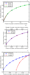
Figure 3. Results for the simulations of Model 2 giving changes in the activity in the modules (normalized by the maximum activity) with changes in inter-modular synaptic connections.
A, comparison between activity and decreases in the fraction of inter-modular synaptic connections (Fext) for three modules (results for each of the three modules indicated by open squares, open circles and open triangles, respectively). Note that there is very little difference in the changes in the normalized activity with changes in the inter-modular extent of synaptic connections for any of the modules. The intra-modular synaptic connectivity fraction F was kept at 0.30 for each module and the coupling strength is S = 0.00075. The number of neurons in each module was 3267 (99×33). B, comparison between normalized activity in the modules and decreases in the fraction of inter-modular synaptic connections (Fext) between them on the one hand (open squares) and activity in the modules and decreases in the strength of synaptic connections on the other (open circles). This is a two modular case, with the results for just one module shown as the other one had identical activity; similar results were obtained for the three module case. The intra-modular synaptic connectivity F was kept at 0.2 and the coupling strength is S = 0.0015. The number of neurons in each module was 2450 (70×35). Note that for the values given in the abscissa there is no difference between changing the extent of inter-modular synaptic connections and changing the strength of the connections. C, comparison between normalized activity in the modules and decreases in the fraction of inter-modular synaptic connections (Fext) between them for the case when the intra-modular extent of synaptic connections was kept at 0.20 (open squares) and the case in which the extent of both inter-modular and intra-modular synaptic connectivity was reduced to the same extent (open circles); S = 0.0015 The number of neurons in each module was 2450 (70×35).
Figure 4
Figure 4. Histogram of the average percentage decreases in CMRglc(ox) determined in rat and primate brains under the experimental conditions given in the abscissa.
CMRglc(ox) is proportional to network activity (Hyder et al.,2013). For primate (blue bars) these are from left to right: lesion of the nucleus of Meynert; the thalamic intralaminar nuclei (Thal); the nucleus of Meynert plus the thalamic nuclei (Mey+Thal); and under the anaesthetic propofol (Prof). For the rat (grey bars) these are from left to right: lesion of the nucleus basilis magnocellularis (NBM); the thalamic intralaminar nuclei (Thal); the NBM plus the thalamic nuclei (NBM+Thal); under the anaesthetic pentobarbital (pent); slices of cortex (slice); and lesion of the parietal cortex (Parietal). The percentages given in this histogram are from Tables S2 to S5.

Similar articles

Cited by

References

    1. Goldman-Rakic PS (1994) Working memory dysfunction in schizophrenia. J Neuropsychiatry Clin Neurosci 6: 348–357. - PubMed
    1. Lee MG, Hassani OK, Alonso A, Jones BE (2005) Cholinergic basal forebrain neurons burst with theta during waking and paradoxical sleep. J Neurosci 25: 4365–4369. - PMC - PubMed
    1. Silver H, Feldman P, Bilker W, Gur RC (2003) Working memory deficit as a core neuropsychological dysfunction in schizophrenia. Am J Psychiatry 160: 1809–1816. - PubMed
    1. Petrides M, Pandya DN (2002) Comparative cytoarchitectonic analysis of the human and the macaque ventrolateral prefrontal cortex and corticocortical connection patterns in the monkey. Eur J Neurosci 16: 291–310. - PubMed
    1. Schmahmann JD, Pandya DN, Wang R, Dai G, D’Arceuil HE, et al. (2007) Association fibre pathways of the brain: parallel observations from diffusion spectrum imaging and autoradiography. Brain 130: 630–653. - PubMed

Publication types