. 2013 Apr 10;8(4):e60538.
doi: 10.1371/journal.pone.0060538. Print 2013.

Purification, gene cloning, and biochemical characterization of a β-glucosidase capable of hydrolyzing sesaminol triglucoside from Paenibacillus sp. KB0549

Affiliations

Purification, gene cloning, and biochemical characterization of a β-glucosidase capable of hydrolyzing sesaminol triglucoside from Paenibacillus sp. KB0549

Arun Nair et al. PLoS One. .

Abstract

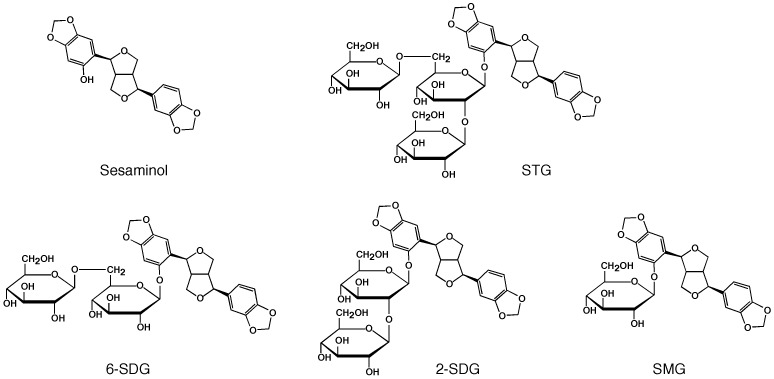
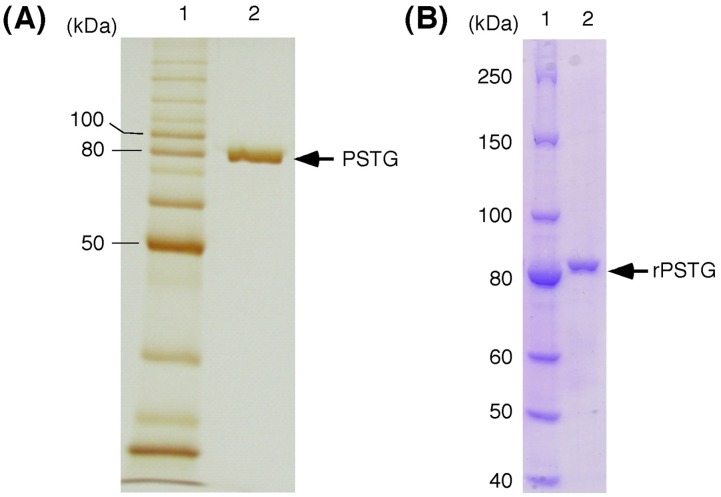
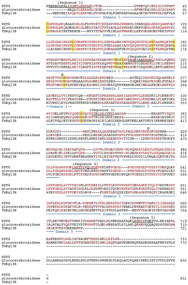
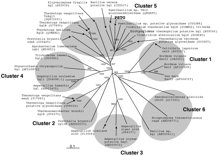
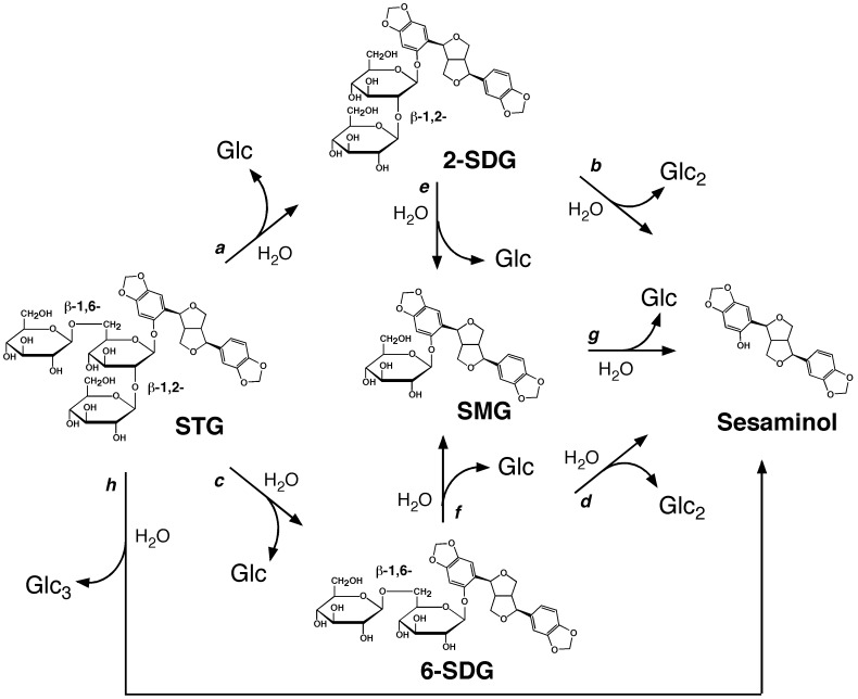
The triglucoside of sesaminol, i.e., 2,6-O-di(β-D-glucopyranosyl)-β-D- glucopyranosylsesaminol (STG), occurs abundantly in sesame seeds and sesame oil cake and serves as an inexpensive source for the industrial production of sesaminol, an anti-oxidant that displays a number of bioactivities beneficial to human health. However, STG has been shown to be highly resistant to the action of β-glucosidases, in part due to its branched-chain glycon structure, and these circumstances hampered the efficient utilization of STG. We found that a strain (KB0549) of the genus Paenibacillus produced a novel enzyme capable of efficiently hydrolyzing STG. This enzyme, termed PSTG, was a tetrameric protein consisting of identical subunits with an approximate molecular mass of 80 kDa. The PSTG gene was cloned on the basis of the partial amino acid sequences of the purified enzyme. Sequence comparison showed that the enzyme belonged to the glycoside hydrolase family 3, with significant similarities to the Paenibacillus glucocerebrosidase (63% identity) and to Bgl3B of Thermotoga neapolitana (37% identity). The recombinant enzyme (rPSTG) was highly specific for β-glucosidic linkage, and k cat and k cat/K m values for the rPSTG-catalyzed hydrolysis of p-nitrophenyl-β-glucopyraniside at 37°C and pH 6.5 were 44 s(-1) and 426 s(-1) mM(-1), respectively. The specificity analyses also revealed that the enzyme acted more efficiently on sophorose than on cellobiose and gentiobiose. Thus, rPSTG is the first example of a β-glucosidase with higher reactivity for β-1,2-glucosidic linkage than for β-1,4- and β-1,6-glucosidic linkages, as far as could be ascertained. This unique specificity is, at least in part, responsible for the enzyme's ability to efficiently decompose STG.


Conflict of interest statement

Competing Interests: The authors here declare the following competing interests, and these do not alter their adherence to all the PLOS ONE policies on sharing data and materials. (1) The following authors are/were employed by Kiyomoto Co. Ltd.: Arun Nair, Ayano Omura, and Kunio Kiyomoyto. (2) There is a granted patent that is related to this paper: JP483923 (inventors, Arun Nair, Kunio Kiyomoyto, Toru Nakayama).

Figures

Figure 1
Figure 1. Structures of sesaminol-related glucosides.
STG, 2,6-O-di(β-D-glucopyranosyl)-β-D-glucopyranosylsesaminol; 2-SDG, 2-O-(β-D -glucopyranosyl)-β-D-glucopyranosylsesaminol; 6-SDG, 6-O-(β-D -glucopyranosyl)-β-D-glucopyranosylsesaminol, and SMG, β-D -glucopyranosylsesaminol.
Figure 2
Figure 2. Reversed-phase HPLC analysis of STG hydrolysis with the crude extract of the KB0549 cells.
The reaction was carried out using method I (see Enzyme assays; final protein concentration, 0.22 mg/ml). Chromatogram A represents that of zero time of the reaction and chromatograms B and C are those for 1 and 3 h after the initiation of the reaction, respectively. Peak a, STG; peak b, 6-SDG; peak c, 2-SDG; peak d, SMG; and peak e, sesaminol.
Figure 3
Figure 3. SDS-PAGE analysis of purified enzymes [(A), PSTG; (B), rPSTG].
Lane 1, molecular mass markers; lane 2, purified enzyme.
Figure 4
Figure 4. Alignment of the deduced amino acid sequence of PSTG protein with those of enzymes (TS12 glucocerebrosidase and TnBgl3B β-glucosidase [28]) belonging to the GH3 family.
Amino acid residues identical to those of PSTG are shown in red. Peptides identified from purified PSTG are underlined (sequences 1–5). The putative catalytic residues of PSTG, Asp233 and Glu421, corresponding to those identified in TS12 glucocerebrosidase by affinity labeling studies and in TnBgl3B by X-ray crystallography , are shown with open circles above the PSTG sequence. Putative sugar-binding amino acid residues at subsite –1 of PSTG and glucocerebrosidase, predicted from the crystal structure of TnBgl3B , are shown with a yellow background. The blue underlining below the TnBgl3B sequence indicates domains 1, 2, and 3 of TnBgl3B identified by X-ray crystallography .
Figure 5
Figure 5. Non-rooted phylogenetic tree of GH3 family glycosidases.
Enzyme names are shown with their DDBJ/EMBL/Genbank accession numbers (parenthesized). The tree was constructed from a CLUSTALW program multiple alignment using a neighbor joining method . Bar = 0.1 amino acid substitutions/site. Numbers indicate bootstrap values greater than 800. Known clusters (clusters 1–6) of GH3 family are shown with gray circles.
Figure 6
Figure 6. Effects of pH and temperature on enzyme activity (A and B) and on the stability (C and D) of rPSTG.
The buffers used were sodium acetate (open circles), potassium phosphate (closed circles), HEPES-NaOH (open rectangles), and glycine-NaOH (closed triangles). The results are presented as the average of three determinations ±SD.
Figure 7
Figure 7. Analysis of the course of sesaminol production from STG catalyzed by rPSTG.
(A) rPSTG (0.15 µM monomer protein) was reacted with 1.15 mM STG (initial concentration, 1.15 mM) at pH 7.0 and 37°C. During the reaction, concentrations of STG (closed circles), 6-STG (open circles), 2-STG (open triangles), SMG (open squares), and sesaminol (closed squares) in the reaction mixture were found by HPLC. Open diamonds show concentrations of STG in the mixture without enzyme. The results are presented as the average of three determinations ±SD. (B) Reversed-phase HPLC analysis of a reaction mixture. Retention times are: STG, 8.52 min; 6-SDG, 10.88 min; SMG, 13.6 min; and sesaminol, 20.92 min. Inset shows co-chromatography of 6-SDG and 2-SDG (11.08 min).
Figure 8
Figure 8. Possible pathways of enzymatic production of sesaminol from STG.

Similar articles

Cited by

References

    1. Fukuda Y, Nagata M, Osawa T, Namiki M (1986) Contribution of lignan analogs to antioxidative activity of refined unroasted sesame seed oil. Journal of the American Oil Chemists’ Society 63: 1027–1031.
    1. Kumazawa S, Koike M, Usui Y, Nakayama T, Fukuda Y (2003) Isolation of sesaminols as antioxidative components from roasted sesame seed oil. Journal of Oleo Science 52: 303–307.
    1. Kang MH, Katsuzaki H, Osawa T (1998) Inhibition of 2,2′-azobis(2,4-dimethylvaleronitrile)-induced lipid peroxidation by sesaminols. Lipids 33: 1031–1036. - PubMed
    1. Kang MH, Naito M, Sakai K, Uchida K, Osawa T (2000) Mode of action of sesame lignans in protecting low-density lipoprotein against oxidative damage in vitro . Life Sciences 66: 161–171. - PubMed
    1. Miyahara Y, Hibasami H, Katsuzaki H, Imai K, Osawa T, et al. (2001) Sesaminol from sesame seed induces apoptosis in human lymphoid leukemia Molt 4B cells. International Journal of Molecular Medicine 7: 485–488. - PubMed

Publication types