. 2013 Apr 12;8(4):e60687.
doi: 10.1371/journal.pone.0060687. Print 2013.

miRNA-27b targets vascular endothelial growth factor C to inhibit tumor progression and angiogenesis in colorectal cancer

Affiliations

miRNA-27b targets vascular endothelial growth factor C to inhibit tumor progression and angiogenesis in colorectal cancer

Jun Ye et al. PLoS One. .

Abstract

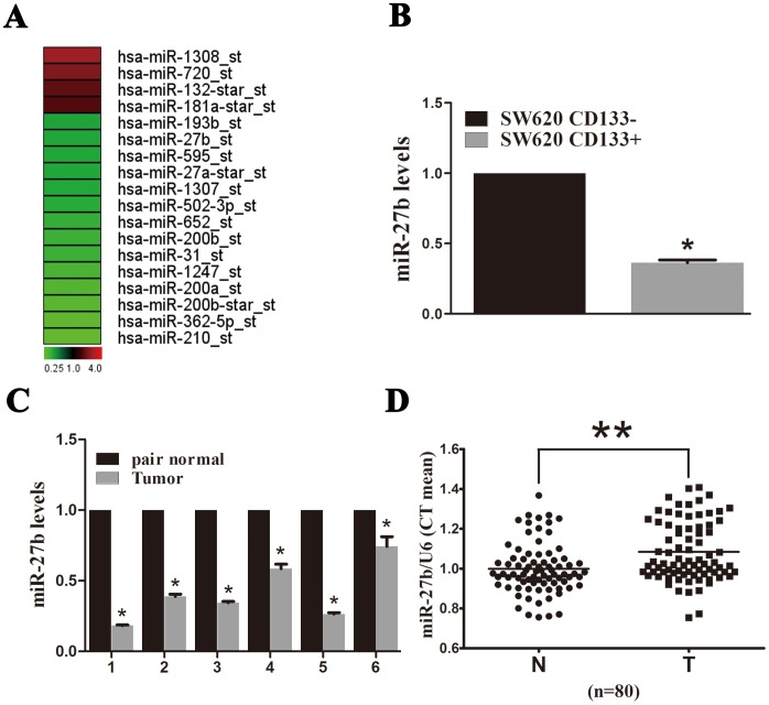
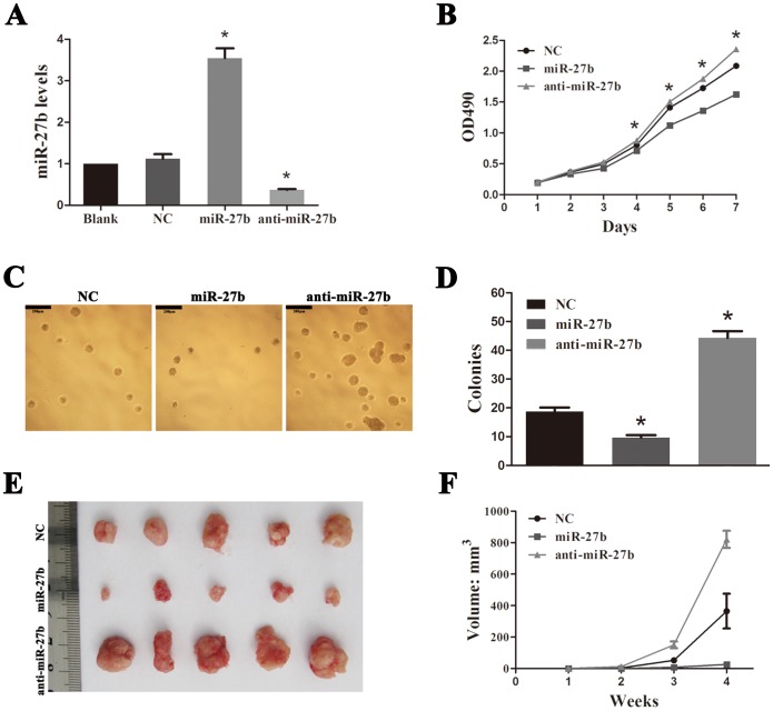
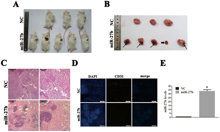
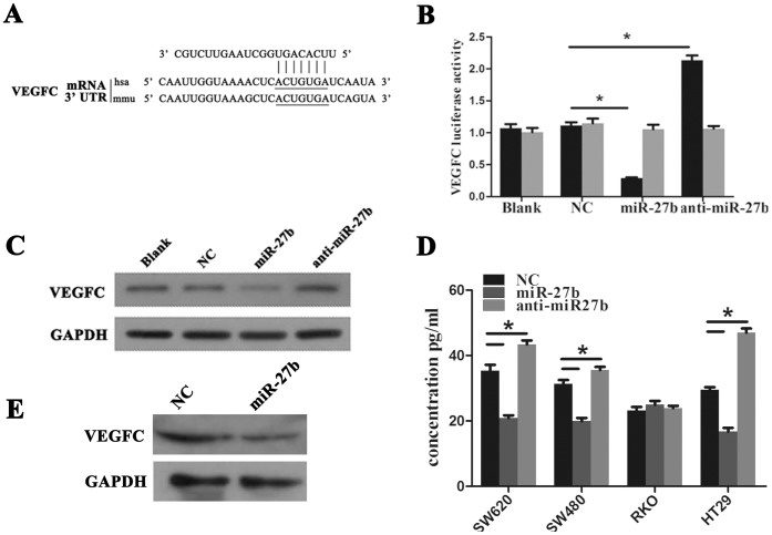
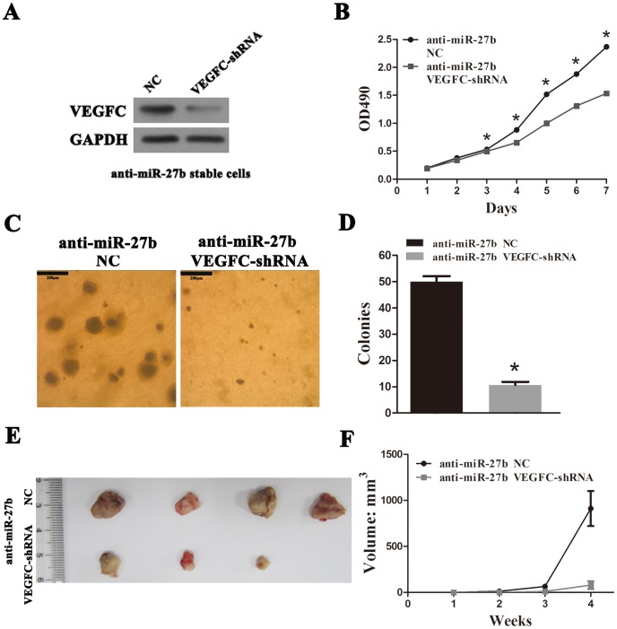
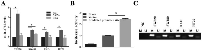
Colorectal cancer (CRC) is one of the most prevalent cancers globally and is one of the leading causes of cancer-related deaths due to therapy resistance and metastasis. Understanding the mechanism underlying colorectal carcinogenesis is essential for the diagnosis and treatment of CRC. microRNAs (miRNAs) can act as either oncogenes or tumor suppressors in many cancers. A tumor suppressor role for miR-27b has recently been reported in neuroblastoma, while no information about miR-27b in CRC is available. In this study, we demonstrated that miR-27b expression is decreased in most CRC tissues and determined that overexpression of miR-27b represses CRC cell proliferation, colony formation and tumor growth in vitro and in vivo. We identified vascular endothelial growth factor C (VEGFC) as a novel target gene of miR-27b and determined that miR-27b functioned as an inhibitor of tumor progression and angiogenesis through targeting VEGFC in CRC. We further determined that DNA hypermethylation of miR-27b CpG islands decreases miR-27b expression. In summary, an anti-tumor role for miR-27b and its novel target VEGFC in vivo could lead to tumor necrosis and provide a rationale for developing miR-27b as a therapeutic agent.


Conflict of interest statement

Competing Interests: The authors have declared that no competing interests exist.

Figures

Figure 1
Figure 1. miR-27b expression in cancer stem cells (CSCs) and colorectal cancer (CRC) tumor tissue.
(A) Differentially expressed miRNA in CD133+ and CD133 cells. Red denotes high and green denotes low levels of expression. (B) miR-27b expression in CD133+ and CD133 cells was assessed by qPCR. The y-axis indicates fold change. (C) miR-27b expression assessed by qPCR in fresh CRC tissues compared to adjacent normal tissues from six patients. The y-axis indicates fold change. (D) miR-27b expression assessed by qPCR in 80 paired paraffin-embedded CRC and adjacent normal tissues. miR-27b levels were normalized to U6 and expressed in terms of the threshold cycle (Ct) ratio. Error bars represent the means ± SEM, *P<0.05, **P<0.01.
Figure 2
Figure 2. miR-27b inhibits tumor growth in colorectal cancer (CRC).
(A) miR-27b expression in negative control (NC), miR-27b and anti-miR-27b SW620 cell lines were assessed by qPCR. The y-axis indicates fold change. (B) Cell proliferation rate detected by measuring the absorbance at 490 nm in an MTS assay. (C and D) Results of a soft-agar colony assay. Colonies were visualized by microscopy after 2 weeks of incubation. Colonies containing >20 cells were counted. Scale bars = 200 µm. (E) Results from a tumorigenesis assay. A representative image of xenograft tumors in nude mice injected subcutaneously with 1×10⁶ CRC cells. (F) Comparison of xenograft formation in vivo. Tumor volumes were measured each week. Error bars represent the means ± SEM, *P<0.05.
Figure 3
Figure 3. miR-27b has anti-tumor and angiogenesis effects in vivo.
(A and B) Colorectal cancer (CRC) bearing NOD/SCID mice were intratumorally injected with cholesterol-conjugated negative control (NC) or miR-27b mimics. Scabs were observed in four tumors from the miR-27b group (fine arrow). One tumor from the miR-27b group disappeared completely with only a scab remaining (thick arrow). (C) Hematoxylin and eosin (HE) staining of xenograft tissues showing necrotic areas in the miR-27b group. (D) A representative immunofluorescence assay showing CD31 protein in xenograft tissues from NC and miR-27b (n = 3). Scale bars = 200 µm. (E) miR-27b expression in xenografts from NC and miR-27b mimics was assessed by qPCR. Error bars represent the means ± SEM, *P<0.05.
Figure 4
Figure 4. Vascular endothelial growth factor C (VEGFC) is a novel target of miR-27b in colorectal cancer (CRC).
(A) VEGFC is predicted as a novel target of miR-27b. (B) 293T cells were co-transfected with empty pmirGLO Dual-Luciferase reporter plasmids or VEGFC 3′UTR firefly luciferase reporter plasmids and pRL-TK-luciferase plasmids, together with miR-27b mimics or anti-miR-27b. After 48 h, firefly luciferase activity was measured and normalized to that of Renilla luciferase. (C) CRC cells were transfected with NC, miR-27b or anti-miR-27b mimics and expression of VEGFC was detected by western blotting. (D) CRC cells were transfected with NC, miR-27b or anti-miR-27b mimics and VEGFC in culture medium was detected by ELISA. (E) VEGFC protein in xenografts from negative control (NC) and miR-27b mimics was detected by western blotting. Error bars represent the means ± SEM, *P<0.05.
Figure 5
Figure 5. Vascular endothelial growth factor C (VEGFC) plays a tumor-promoting role in colorectal cancer (CRC).
(A) VEGFC knockdown in anti-miR-27b stable cells was confirmed by western blotting. (B) Cell proliferation rate was determined by measuring the absorbance at 490 nm in a MTS assay. (C and D) Results of a soft-agar colony assay. Colonies were visualized by microscopy after 2 weeks of incubation. Colonies containing >20 cells were counted. Scale bars = 200 µm. (E) Results of a tumorigenesis assay. A representative image of xenograft tumors in nude mice subcutaneously injected with 1×10⁶ CRC cells. (F) Comparison of xenograft formation in vivo. Tumor volumes were measured each week. Error bars represent the means ± SEM, *P<0.05.
Figure 6
Figure 6. DNA hypermethylation is associated with decreased miR-27b expression.
(A) The levels of miR-27b expression in colorectal cancer (CRC) cell lines were determined by qPCR after treatment with 5AZA or TSA for 3 days. (B) Results of luciferase activity assays following transfection with the predicted miR-27b promoter normalized to pRL-TK Renilla luciferase. (C) MSP analysis of the miR-27b CpG island in a set of CRC cell lines. Bands in the ‘M’ lanes are PCR products obtained using methylation-specific primers and those in the ‘U’ lanes are products obtained using unmethylated-specific primers.

Similar articles

Cited by

References

    1. Ferlay J, Shin HR, Bray F, Forman D, Mathers C, et al. (2010) Estimates of worldwide burden of cancer in 2008: GLOBOCAN 2008. Int J Cancer 127: 2893–2917. - PubMed
    1. Vo DM, Julien LA, Thorson AG (2010) Current controversies in colon and rectal cancer. Minerva Chir 65: 677–693. - PubMed
    1. Greenlee RT, Hill-Harmon MB, Murray T, Thun M (2001) Cancer statistics, 2001. CA Cancer J Clin 51: 15–36. - PubMed
    1. Mueller MM, Fusenig NE (2004) Friends or foes - bipolar effects of the tumour stroma in cancer. Nature Reviews Cancer 4: 839–849. - PubMed
    1. Balkwill F (2004) Cancer and the chemokine network. Nature Reviews Cancer 4: 540–550. - PubMed

Publication types

MeSH terms