. 2013 Apr 9;8(4):e60717.
doi: 10.1371/journal.pone.0060717. Print 2013.

Klebsiella phage vB_KleM-RaK2 - a giant singleton virus of the family Myoviridae

Affiliations

Klebsiella phage vB_KleM-RaK2 - a giant singleton virus of the family Myoviridae

Eugenijus Simoliūnas et al. PLoS One. .

Erratum in

  • PLoS One. 2013;8(12). doi:10.1371/annotation/a1d15675-2942-41ba-92f4-3dad6bc6cac6

Abstract

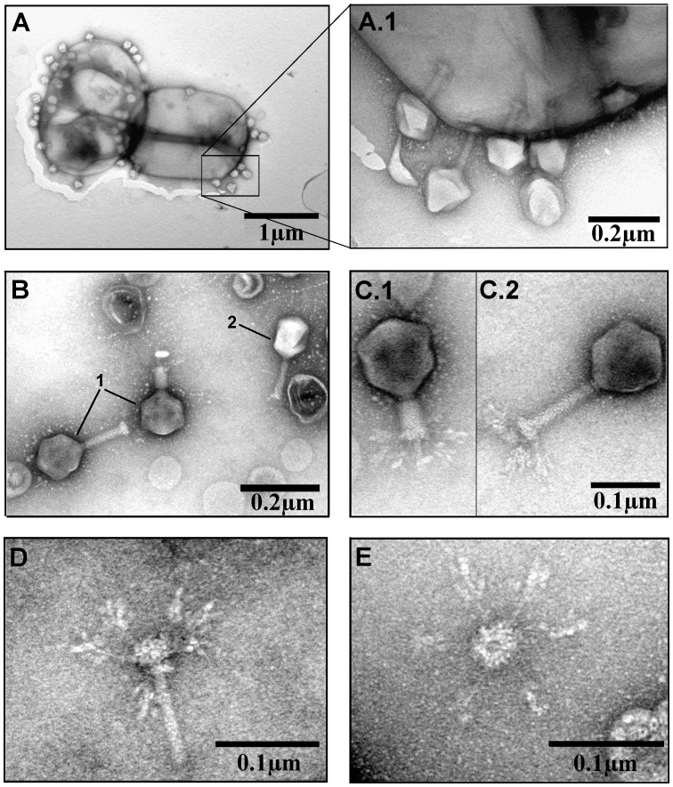
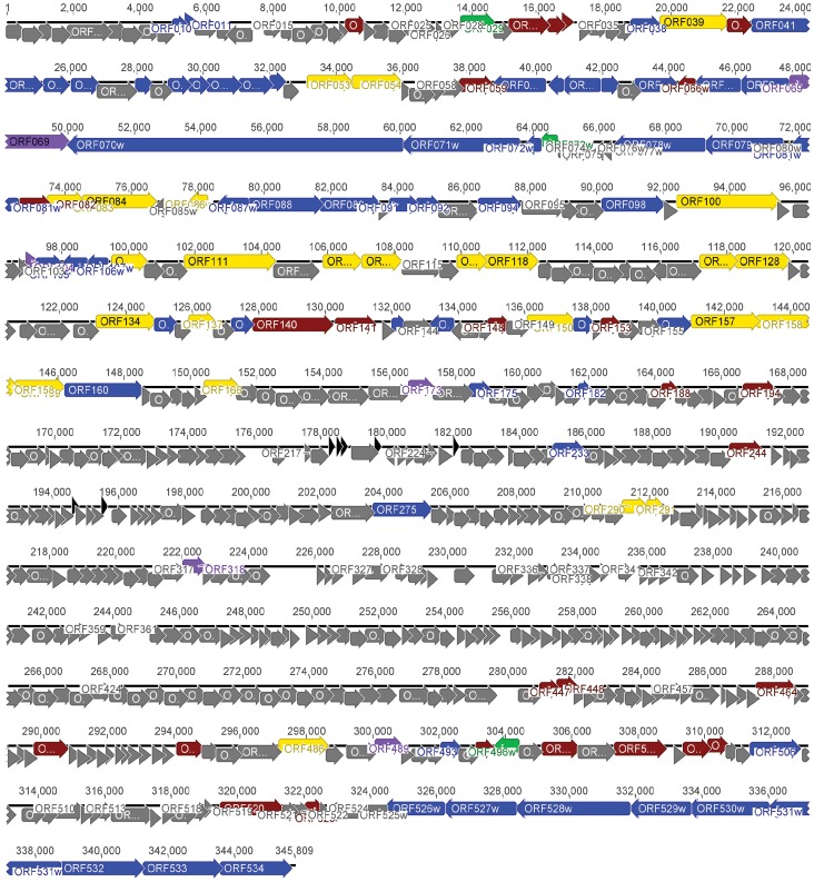
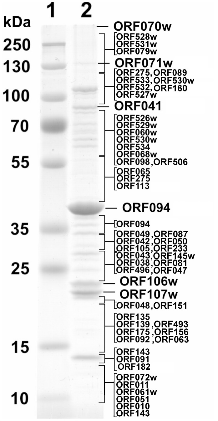
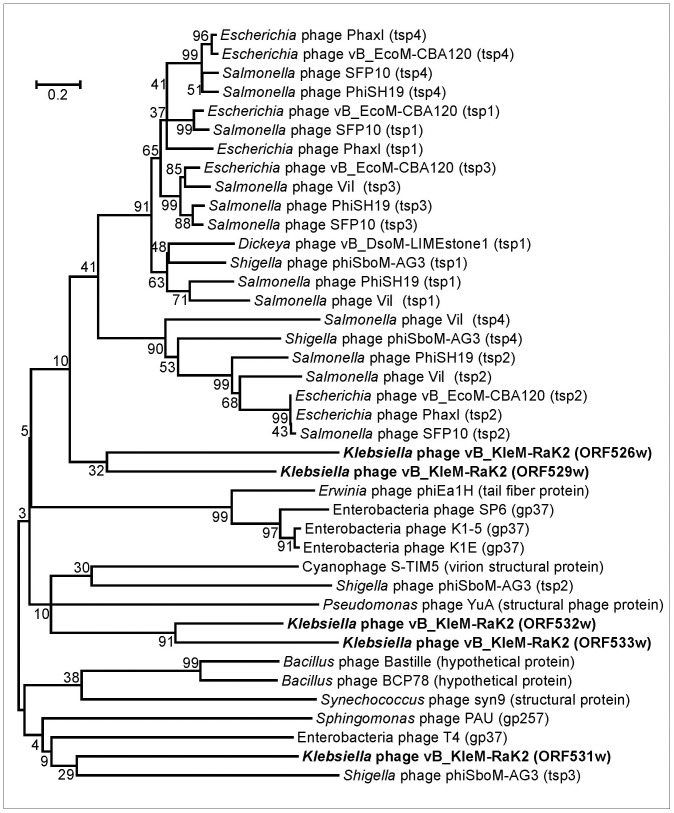
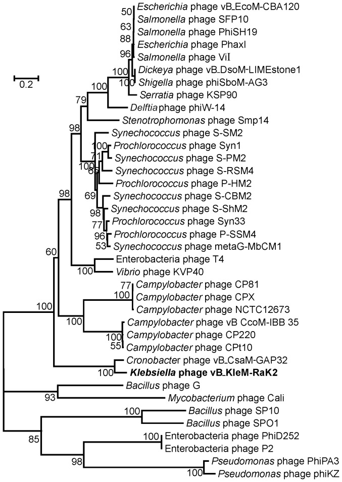
At 346 kbp in size, the genome of a jumbo bacteriophage vB_KleM-RaK2 (RaK2) is the largest Klebsiella infecting myovirus genome sequenced to date. In total, 272 out of 534 RaK2 ORFs lack detectable database homologues. Based on the similarity to biologically defined proteins and/or MS/MS analysis, 117 of RaK2 ORFs were given a functional annotation, including 28 RaK2 ORFs coding for structural proteins that have no reliable homologues to annotated structural proteins in other organisms. The electron micrographs revealed elaborate spike-like structures on the tail fibers of Rak2, suggesting that this phage is an atypical myovirus. While head and tail proteins of RaK2 are mostly myoviridae-related, the bioinformatics analysis indicate that tail fibers/spikes of this phage are formed from podovirus-like peptides predominantly. Overall, these results provide evidence that bacteriophage RaK2 differs profoundly from previously studied viruses of the Myoviridae family.


Conflict of interest statement

Competing Interests: The authors have declared that no competing interests exist.

Figures

Figure 1
Figure 1. Electron micrographs of Klebsiella phage RaK2.
(A and A.1) Phage RaK2 particles adsorbed to the surface of Klebsiella sp. KV-3 cells. (B) Purified phage RaK2 particles (1) and one particle of phage T4 (2). (C) RaK2 particle with contracted (C.1) and extended (C.2) tail. (D) Inner tail tube with baseplate and baseplate-associated ramified tail fiber structures. (E) Baseplate with six long tail fibers.
Figure 2
Figure 2. Single-step growth curves of bacteriophage RaK2 at 30°C.
Shown are PFU per infected Klebsiella sp. KV-3 cell in chloroform-treated cultures (filled circle) and untreated cultures (circle) at different time points. Each point represents the mean of three individual experiments.
Figure 3
Figure 3. Growth of RaK2 plaques at 30°C.
Numbers above indicate days of incubation.
Figure 4
Figure 4. Functional genome map of bacteriophage RaK2.
The coding capacity of the RaK genome is shown. Functions are assigned according to the characterized ORFs in NCBI database and/or MS/MS analysis. The colour code is as follows: yellow – DNA replication, recombination, repair and packaging; brown – transcription, translation, nucleotide metabolism; blue – structural proteins; purple – chaperones/assembly; green – lysis, host or phage interactions; grey – ORFs of unknown function; black – tRNA and pseudo-tRNA.
Figure 5
Figure 5. SDS-PAGE of virion proteins of RaK2.
Lanes: 1– molecular mass marker Page Ruler™ prestained Protein Ladder Plus (Thermofisher), 2– phage RaK2. Relative migrations of MW marker proteins are indicated on the left. Proteins identified by MS/MS are indicated on the right.
Figure 6
Figure 6. Neighbor-joining tree based on the alignment of the tail spike/tail fiber proteins from various phage genomes.
The bootstrap values indicated.
Figure 7
Figure 7. Neighbor-joining tree analysis based on the alignment of the amino acid sequences of the major capsid proteins from various myoviruses.
The numbers at the nodes indicate the bootstrap probabilities.
Figure 8
Figure 8. Phylogenetic analysis.
Relationships of (A) DNA ligase; (B) DNA polymerase; (C) terminase large subunit; (D) thymidylate synthase across diverse phage types. The bootstrap values are indicated.

Similar articles

Cited by

References

    1. Wommack KE, Colwell RR (2000) Virioplankton: viruses in aquatic ecosystems. Microbiol Mol Biol Rev 64: 69–114. - PMC - PubMed
    1. Hendrix RW (2002) Bacteriophages: evolution of the majority. Theor Popul Biol 61: 471–480. - PubMed
    1. Thomas JA, Weintraub ST, Pakala K, Server P, Hardies SC (2010) Proteome of the large Pseudomonas myovirus 201φ2–1. Mol Cell Proteomics 9: 940–951. - PMC - PubMed
    1. Ackerman H-W (2011) Bacteriophage taxonomy. Cambridge Publishing, Microbiology. Australia.
    1. Comeau AM, Tremblay D, Moineau S, Rattei T, Kushkina AI, et al. (2012) Phage morphology recapitulates phylogeny: the comparative genomics of a new group of myoviruses. PLoS One 7: e40102. - PMC - PubMed

Publication types

Associated data