Comparative Study
. 2013 Apr 11;8(4):e60734.
doi: 10.1371/journal.pone.0060734. Print 2013.

Structural and functional role of INI1 and LEDGF in the HIV-1 preintegration complex

Affiliations
Comparative Study

Structural and functional role of INI1 and LEDGF in the HIV-1 preintegration complex

Benoit Maillot et al. PLoS One. .

Abstract

Integration of the HIV-1 cDNA into the human genome is catalyzed by the viral integrase (IN) protein. Several studies have shown the importance of cellular cofactors that interact with integrase and affect viral integration and infectivity. In this study, we produced a stable complex between HIV-1 integrase, viral U5 DNA, the cellular cofactor LEDGF/p75 and the integrase binding domain of INI1 (INI1-IBD), a subunit of the SWI/SNF chromatin remodeling factor. The stoichiometry of the IN/LEDGF/INI1-IBD/DNA complex components was found to be 4/2/2/2 by mass spectrometry and Fluorescence Correlation Spectroscopy. Functional assays showed that INI1-IBD inhibits the 3' processing reaction but does not interfere with specific viral DNA binding. Integration assays demonstrate that INI1-IBD decreases the amount of integration events but inhibits by-product formation such as donor/donor or linear full site integration molecules. Cryo-electron microscopy locates INI1-IBD within the cellular DNA binding site of the IN/LEDGF complex, constraining the highly flexible integrase in a stable conformation. Taken together, our results suggest that INI1 could stabilize the PIC in the host cell, by maintaining integrase in a stable constrained conformation which prevents non-specific interactions and auto integration on the route to its integration site within nucleosomes, while LEDGF organizes and stabilizes an active integrase tetramer suitable for specific vDNA integration. Moreover, our results provide the basis for a novel type of integrase inhibitor (conformational inhibitor) representing a potential new strategy for use in human therapy.


Conflict of interest statement

Competing Interests: The authors have read the journal’s policy and have the following interest: Author Alexis Nazabal is affiliated to the commercial company CovalX AG. This does not alter the authors’ adherence to all the PLOS ONE policies on sharing data and materials.

Figures

Figure 1
Figure 1. In vitro functional tests.
A–B: Determination by fluorescence anisotropy of the dissociation constant of viral DNA to IN/LEGDF and IN/LEDGF/INI1-IBD. A: Titration curve of viral DNA by the IN/LEDGF complex in the absence (square, continuous line) or in the presence of an excess of non-fluorescent viral DNA (circles, dotted line) and non-fluorescent non-specific DNA (triangles, dashed line). B: Same as A for the IN/LEDGF/INI1-IBD complex. Lines correspond to the fits of equation 8 in methods S1 to the data. C: 3′ processing reaction followed by fluorescence anisotropy. The fraction of released GT as a function of time is shown in blue for the IN/LEDGF complex and in red for the IN/LEDGF/INI1-IBD complex.
Figure 2
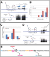
Figure 2. Comparative analysis of the in vitro concerted integration catalyzed by HIV-1 IN, HIV-1 IN/LEDGF and HIV-1 IN/LEDGF/INI1-IBD complex.
A. In vitro integration profiles of HIV-1 IN and HIV-1 IN/LEDGF complexes. Concerted integration assay was performed using 0.1 to 12.5µM IN or IN/LEDGF complex (concentration are normalized to IN monomers), 150 ng of acceptor plasmid, 15 ng of radioactively labelled and processed donor DNA. The reaction products were loaded on 1% agarose gel. The position and the structure of the different products obtained after half-site (HSI), full-site (FSI) and donor/donor (d/d) integration are reported. B. Full site integration activity. The circular FSI products obtained in (A) were quantified by cloning in bacteria and reported as the number of ampicillin-, kanamycin- and tetracycline-resistant selected clones . C. Structure of the integration products. One hundred circular FSI integration products were isolated in condition where the concerted integration assay was performed with 12.5 µM of IN or IN/LEDGF complexes and were sequenced as reported in the materials and methods section. The number of correct 5 bp duplication, deletions and other structures are reported as percentages. All values reported are the mean ± standard deviation (error bars) of three independent sets of experiments. D. Effect of INI1 on the integration profile. The assay was performed and reported as in (A) using 12.5µM IN, IN/LEDGF or IN/LEDGF/INI1-IBD complex (in IN monomers). A quantification of the gel showing the strong decrease in linear fsi and dd product is shown in figure S8. E Proposed equilibrium between the different complexes involved. In the presence of tDNA, INI1 is slowly displaced from the IN/LEDGF/INI1-IBD/vDNA complex and the integration reaction can occur to produce specific integration products.
Figure 3
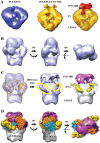
Figure 3. Cryo-EM structure.
A. Negatively stained structures of the IN/LEDGF (blue) and IN/LEDGF/INI1-IBD (gold) complexes. The difference between the two maps representing the position of INI1-IBD is in red. B. Cryo-EM structure of the IN/LEDGF/INI1-IBD/DNA complex represented by three perpendicular views. C. Difference map calculated by subtracting the density corresponding to the fitted atomic models of IN and LEDGF from the IN/LEDGF/INI1-IBD/DNA cryo-EM map. The differences, corresponding respectively to INI1-IBD, DNA and LEDGF, are represented in violet, yellow and grey. D. Three perpendicular views of the IN/LEDGF/INI1-IBD/DNA complex: INI1-IBD is in violet, LEDGF in grey and viral DNA in yellow. The fitted IN model is represented as molecular surface. For both IN dimers, the two monomers are in blue and gold. The two IN dimers (light and dark) are related by a twofold symmetry.
Figure 4
Figure 4. Superimposition of the IN/LEDGF/DNA and IN/LEDGF/INI1-IBD/DNA structures.
A: Superimposition of the IN/LEDGF/DNA (grey) and IN/LEDGF/INI1-IBD/DNA (blue) structures. B: DNA fitting. The spikes in the two structures unambigously revealed the DNA position. The DNA (yellow) is fitted in the IN/LEDGF/INI1-IBD/DNA map (mesh representation) with a 40° rotation from its position in the IN/LEDGF/DNA map (surface representation).
Figure 5
Figure 5. Topology of the IN tetramer.
A: View from the INI1-IBD side of the complex. B: View of the IN tetramer after removing the INI1-IBD density. The two IN dimers (1 and 2) are related by a twofold axis perpendicular to the sheet. Each IN monomer (A and B) is represented in a different color (blue and gold). The two monomers related by the two fold symmetry are represented in light and dark color (light blue for A1 and dark blue for A2, light gold for B1 and dark gold for B2). The positions of the IN domains are indicated: Nt for the N-terminal domain, Cc for the catalytic core and Ct for the C-terminal domain.
Figure 6
Figure 6. IN/LEDGF/INI1-IBD/DNA structure.
A. INI1-IBD (pink) sits on the top of the complex and interacts with the C and N termini of the two IN monomers B. An extension of INI1-IBD interacts with the C-terminus of the two monomers A. B. A 90° view sliced as shown by the dotted square in figure A. C and D: two 90° views showing clearly the IN interacting domains with INI1.
Figure 7
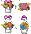
Figure 7. Structure-function analysis.
A. IN/LEDGF/vDNA structure in the 3′ processing conformation. B. In the IN/LEDGF/INI1-IBD/vDNA complex, INI1-IBD caps the surface of the complex and blocks IN in an intermediate conformation, as shown by the position of the viral DNA (yellow). C. IN/LEDGF/vDNA/tDNA structure in the strand transfer conformation after release of INI1 and binding to the target DNA (red).

Similar articles

Cited by

References

    1. Briones MS, Dobard CW, Chow SA (2010) Role of human immunodeficiency virus type 1 integrase in uncoating of the viral core. JVirol 84: 5181–5190. - PMC - PubMed
    1. Wu X, Liu H, Xiao H, Conway JA, Hehl E, et al. (1999) Human immunodeficiency virus type 1 integrase protein promotes reverse transcription through specific interactions with the nucleoprotein reverse transcription complex. JVirol 73: 2126–2135. - PMC - PubMed
    1. Ao Z, Danappa JK, Wang B, Zheng Y, Kung S, et al. (2010) Importin alpha3 interacts with HIV-1 integrase and contributes to HIV-1 nuclear import and replication. J Virol 84: 8650–8663. - PMC - PubMed
    1. Hendrix J, Gijsbers R, De RJ, Voet A, Hotta JI, et al. (2011) The transcriptional co-activator LEDGF/p75 displays a dynamic scan-and-lock mechanism for chromatin tethering. Nucleic Acids Res 39: 1310–1325. - PMC - PubMed
    1. Chiu TK, Davies DR (2004) Structure and function of HIV-1 integrase. CurrTopMedChem 4: 965–977. - PubMed

Publication types

MeSH terms