. 2013 Apr 8;8(4):e60817.
doi: 10.1371/journal.pone.0060817. Print 2013.

Minocycline suppresses interleukine-6, its receptor system and signaling pathways and impairs migration, invasion and adhesion capacity of ovarian cancer cells: in vitro and in vivo studies

Affiliations

Minocycline suppresses interleukine-6, its receptor system and signaling pathways and impairs migration, invasion and adhesion capacity of ovarian cancer cells: in vitro and in vivo studies

Parvin Ataie-Kachoie et al. PLoS One. .

Expression of concern in

Abstract

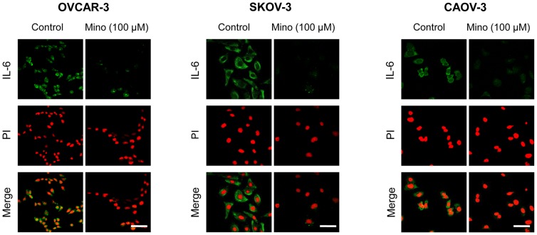
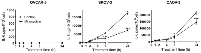
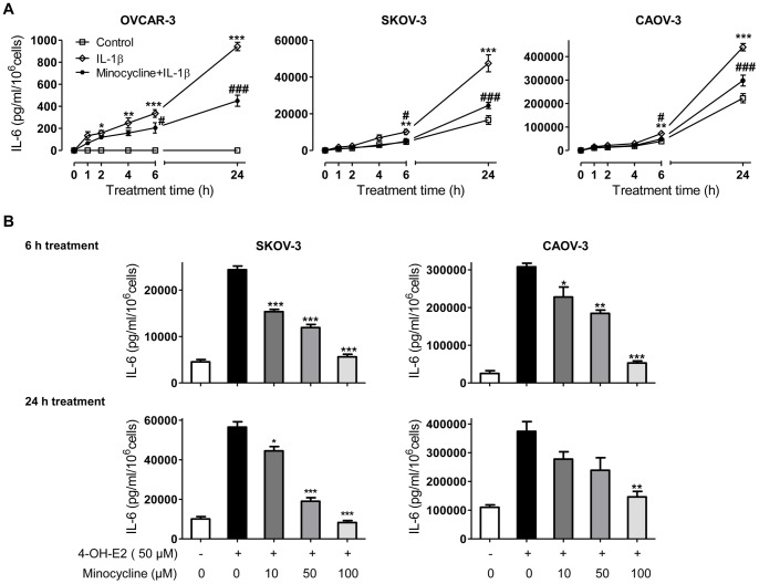
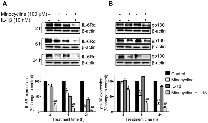
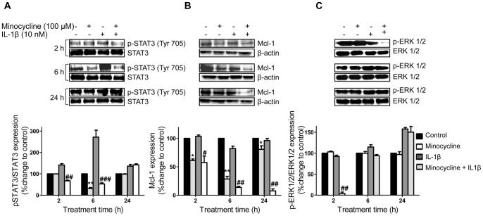
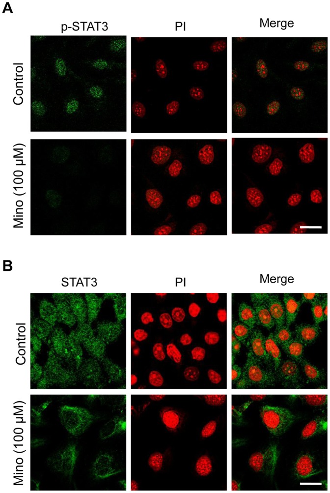
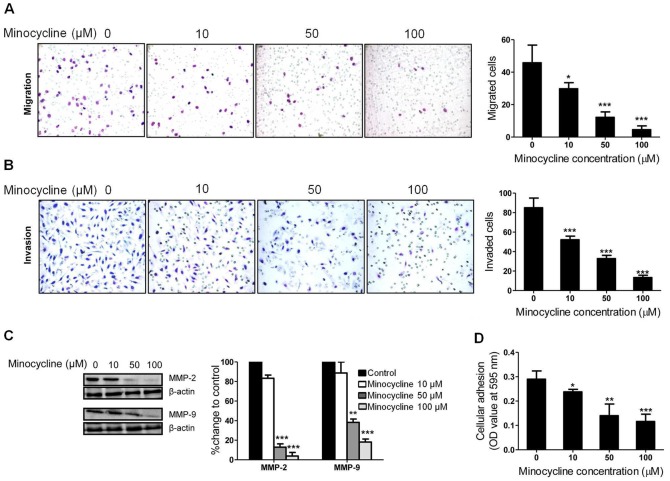
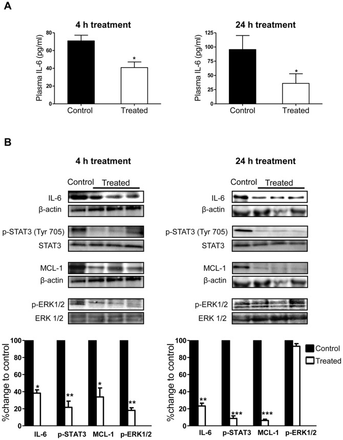
Interleukin (IL)-6 has been shown to be a major contributing factor in growth and progression of ovarian cancer. The cytokine exerts pro-tumorigenic activity through activation of several signaling pathways in particular signal transducer and activator of transcription (STAT3) and extracellular signal-regulated kinase (ERK)1/2. Hence, targeting IL-6 is becoming increasingly attractive as a treatment option in ovarian cancer. Here, we investigated the effects of minocycline on IL-6 and its signaling pathways in ovarian cancer. In vitro, minocycline was found to significantly suppress both constitutive and IL-1β or 4-hydroxyestradiol (4-OH-E2)-stimulated IL-6 expression in human ovarian cancer cells; OVCAR-3, SKOV-3 and CAOV-3. Moreover, minocycline down-regulated two major components of IL-6 receptor system (IL-6Rα and gp130) and blocked the activation of STAT3 and ERK1/2 pathways leading to suppression of the downstream product MCL-1. In female nude mice bearing intraperitoneal OVCAR-3 tumors, acute administration (4 and 24 h) of minocycline (30 mg/kg) led to suppression of IL-6. Even single dose of minocycline was effective at significantly lowering plasma and tumor IL-6 levels. In line with this, tumoral expression of p-STAT3, p-ERK1/2 and MCL-1 were decreased in minocycline-treated mice. Evaluation of the functional implication of minocycline on metastatic activity revealed the capacity of minocycline to inhibit cellular migration, invasion and adhesion associated with down-regulation of matrix metalloproteinases (MMP)-2 and 9. Thus, the data suggest a potential role for minocycline in suppressing IL-6 expression and activity. These effects may prove to be an important attribute to the upcoming clinical trials of minocycline in ovarian cancer.


Conflict of interest statement

Competing Interests: The authors have declared that no competing interests exist.

Figures

Figure 1
Figure 1. Minocycline decreases constitutive expression of IL-6 in ovarian cancer cell lines.
Representative confocal images of IL-6 (green) in OVCAR-3, SKOV-3 and CAOV-3 cells under control conditions and exposed to minocycline (100 µM) for 24 h. Cells were also stained with propidium iodide (red). Images were obtained at 60× magnification. The scale bars represent 20 µm.
Figure 2
Figure 2. Effect of minocycline on basal IL-6 levels in media bathing ovarian cancer cells.
Minocycline (100 µM) decreased IL-6 expression of SKOV-3 and CAOV-3 cells in a time-dependent pattern as analyzed by ELISA. The values shown are mean ± SD of data from three independent experiments (*p<0.05 and ***p<0.001 vs. control group).
Figure 3
Figure 3. Minocycline inhibits IL-6 surge in stimulated ovarian cancer cell lines.
(A) OVCAR-3, SKOV-3 and CAOV-3 cells were pre-treated with minocycline (100 µM) followed by stimulation with IL-1β (10 ng/ml) for different time points. Levels of IL-6 in the culture media were quantified by sandwich ELISA. Each data represent the mean ± SD from three independent experiments. (*p<0.05, **p<0.01 and ***p<0.001 vs. control group, #p<0.05 and ### p<0.01 vs. IL-1β-stimulated group). (B) OVCAR-3, SKOV-3 and CAOV-3 cells were pre-treated for 1 h with varying concentrations of minocycline (0–100 µM) followed by stimulation with 4-OH-E2 (50 µg/ml) for 6 and 24 h. Media were collected and IL-6 level was analyzed by ELISA. The values shown are mean ± SD of data from three independent experiments (*p<0.05, **p<0.01 and ***p<0.001 vs. 4-OH-E2-stimulated group).
Figure 4
Figure 4. Effects of minocycline on IL-6Rα and gp130 expression in ovarian cancer cells.
To detect expression levels of (A) IL-6Rα or (B) gp130, SKOV-3 cells were either stimulated with IL-1β (10 ng/ml) or non-stimulated, with or without pre-treatment with minocycline (100 µM) for different time points. Cell lysates were analyzed by immunoblotting for IL-6Rα, gp130 and β-actin antibodies. β-actin was the loading control. IL-6Rα and gp130 protein levels were normalized to β-actin and their relative differences with the corresponding controls are shown (*p<0.05 and **p<0.01 vs. control cells, # p<0.05 and## p<0.01 vs. IL-1β treated cells).
Figure 5
Figure 5. Influence of minocycline on p-STAT3, p-ERK1/2 and Mcl-1 expression in ovarian cancer cells.
SKOV-3 cells were treated with minocycline (100 µM) with or without IL-1β (10 ng/ml) stimulation for different time points. The expression levels of (A) p-STAT3, STAT3; (B) Mcl-1 or (C) p-ERK1/2, ERK1/2 were estimated by western blot analysis. Densitometric analysis is expressed as mean ± SD intensity of optical density obtained by three independent experiments (*p<0.05, **p<0.01 and ***p<0.001 vs. control cells, # p<0.05, ## p<0.01 vs. IL-1β treated cells).
Figure 6
Figure 6. Effect of minocycline on STAT3 nuclear translocation.
Confocal immunocytochemistry of (A) p-STAT3 and (B) STAT3 (green) in SKOV-3 cells treated with 100 µM minocycline in comparison with control cells. The nuclei are counterstained with propidium iodide (red). Images were obtained at 60× magnification. The scale bars represent 10 µm.
Figure 7
Figure 7. Minocycline inhibits metastatic capacity of SKOV-3 cells along with down-regulation of MMP-2 and MMP-9.
The effects of minocycline on SKOV-3 (A) cell migration and (B) invasion was measured by Transwell assay. Minocycline was applied at different concentrations (0–100 µM) for 18 h. (C) Expression of MMP-2 and MMP-9 estimated by western blot analysis after 18 h treatment of SKOV-3 cells with varying concentration of minocycline. (D) Cell adhesion following 18 h exposure to varying concentrations of minocycline as described in MATERIAL AND METHODS (*p<0.05, **p<0.01 and ***p<0.001 vs. control).
Figure 8
Figure 8. Suppression of IL-6 and its pathways by minocycline in mice bearing ovarian tumors.
(A) IL-6 levels were determined by ELISA in the plasma of mice bearing i.p. OVCAR-3 tumors, 4 and 24 h post minocycline treatment (single dose 30 mg/kg; i.p.) (B) Tumors harvested at 4 and 24 h were examined by western blot analysis for the expression of IL-6, p-STAT3 (Tyr 705), MCL-1 and p-ERK1/2 protein. (*p<0.05, **p<0.01 and ***p<0.001 vs. control).

References

    1. Vaughan S, Coward JI, Bast RC Jr, Berchuck A, Berek JS, et al. (2011) Rethinking ovarian cancer: recommendations for improving outcomes. Nat Rev Cancer 11: 719–725. - PMC - PubMed
    1. Maccio A, Madeddu C (2012) Inflammation and ovarian cancer. Cytokine 58: 133–147. - PubMed
    1. Coward J, Kulbe H, Chakravarty P, Leader D, Vassileva V, et al. (2011) Interleukin-6 as a Therapeutic Target in Human Ovarian Cancer. Clin Cancer Res 17: 6083–6096. - PMC - PubMed
    1. Guo Y, Xu F, Lu T, Duan Z, Zhang Z (2012) Interleukin-6 signaling pathway in targeted therapy for cancer. Cancer Treat Rev. - PubMed
    1. Gastl G, Plante M (2001) Bioactive interleukin-6 levels in serum and ascites as a prognostic factor in patients with epithelial ovarian cancer. Methods Mol Med 39: 121–123. - PubMed

MeSH terms