A patient with posterior cortical atrophy possesses a novel mutation in the presenilin 1 gene
- PMID: 23593396
- PMCID: PMC3625161
- DOI: 10.1371/journal.pone.0061074
A patient with posterior cortical atrophy possesses a novel mutation in the presenilin 1 gene
Abstract
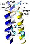
Posterior cortical atrophy is a dementia syndrome with symptoms of cortical visual dysfunction, associated with amyloid plaques and neurofibrillary tangles predominantly affecting visual association cortex. Most patients diagnosed with posterior cortical atrophy will finally develop a typical Alzheimer's disease. However, there are a variety of neuropathological processes, which could lead towards a clinical presentation of posterior cortical atrophy. Mutations in the presenilin 1 gene, affecting the function of γ-secretase, are the most common genetic cause of familial, early-onset Alzheimer's disease. Here we present a patient with a clinical diagnosis of posterior cortical atrophy who harbors a novel Presenilin 1 mutation (I211M). In silico analysis predicts that the mutation could influence the interaction between presenilin 1 and presenilin1 enhancer-2 protein, a protein partner within the γ-secretase complex. These findings along with published literature support the inclusion of posterior cortical atrophy on the Alzheimer's disease spectrum.
Conflict of interest statement
Figures


References
-
- Goldstein MA, Ivanov I, Silverman ME (2011) Posterior cortical atrophy: an exemplar for renovating diagnostic formulation in neuropsychiatry. Compr Psychiatry 52: 326–333. - PubMed
-
- Hof PR, Vogt BA, Bouras C, Morrison JH (1997) Atypical form of Alzheimer's disease with prominent posterior cortical atrophy: a review of lesion distribution and circuit disconnection in cortical visual pathways. Vision Res 37: 3609–3625. - PubMed
-
- Alladi S, Xuereb J, Bak T, Nestor P, Knibb J, et al. (2007) Focal cortical presentations of Alzheimer's disease. Brain 130: 2636–2645. - PubMed
-
- Tang-Wai DF, Graff-Radford NR, Boeve BF, Dickson DW, Parisi J (2004) Clinical, genetic, and neuropathologic characteristics of posterior cortical atrophy. Neurology 63: 1168–1174. - PubMed
Publication types
MeSH terms
Substances
LinkOut - more resources
Full Text Sources
Other Literature Sources

