

Mouse nuclear myosin I knock-out shows interchangeability and redundancy of myosin isoforms in the cell nucleus
- PMID: 23593477
- PMCID: PMC3623870
- DOI: 10.1371/journal.pone.0061406
Mouse nuclear myosin I knock-out shows interchangeability and redundancy of myosin isoforms in the cell nucleus
Abstract
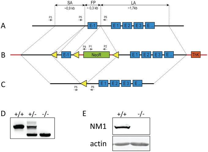
Background: Nuclear myosin I (NM1) is a nuclear isoform of the well-known "cytoplasmic" Myosin 1c protein (Myo1c). Located on the 11(th) chromosome in mice, NM1 results from an alternative start of transcription of the Myo1c gene adding an extra 16 amino acids at the N-terminus. Previous studies revealed its roles in RNA Polymerase I and RNA Polymerase II transcription, chromatin remodeling, and chromosomal movements. Its nuclear localization signal is localized in the middle of the molecule and therefore directs both Myosin 1c isoforms to the nucleus.
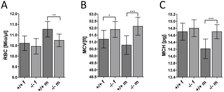
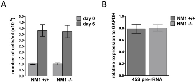
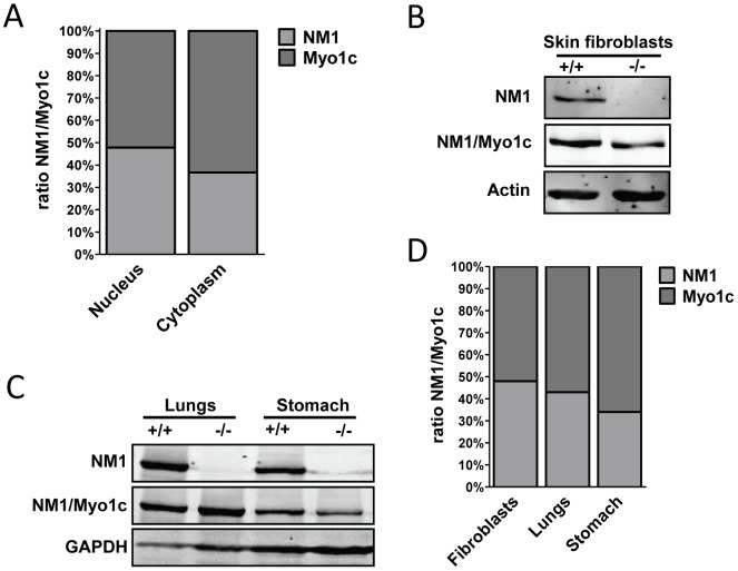
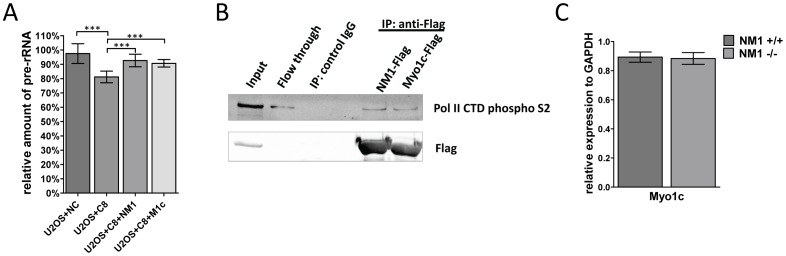
Methodology/principal findings: In order to trace specific functions of the NM1 isoform, we generated mice lacking the NM1 start codon without affecting the cytoplasmic Myo1c protein. Mutant mice were analyzed in a comprehensive phenotypic screen in cooperation with the German Mouse Clinic. Strikingly, no obvious phenotype related to previously described functions has been observed. However, we found minor changes in bone mineral density and the number and size of red blood cells in knock-out mice, which are most probably not related to previously described functions of NM1 in the nucleus. In Myo1c/NM1 depleted U2OS cells, the level of Pol I transcription was restored by overexpression of shRNA-resistant mouse Myo1c. Moreover, we found Myo1c interacting with Pol II. The ratio between Myo1c and NM1 proteins were similar in the nucleus and deletion of NM1 did not cause any compensatory overexpression of Myo1c protein.
Conclusion/significance: We observed that Myo1c can replace NM1 in its nuclear functions. Amount of both proteins is nearly equal and NM1 knock-out does not cause any compensatory overexpression of Myo1c. We therefore suggest that both isoforms can substitute each other in nuclear processes.
Conflict of interest statement
Figures
Similar articles
-
Specific nuclear localizing sequence directs two myosin isoforms to the cell nucleus in calmodulin-sensitive manner.PLoS One. 2012;7(1):e30529. doi: 10.1371/journal.pone.0030529. Epub 2012 Jan 25. PLoS One. 2012. PMID: 22295092 Free PMC article.
-
Nuclear myosin 1 contributes to a chromatin landscape compatible with RNA polymerase II transcription activation.BMC Biol. 2015 Jun 5;13:35. doi: 10.1186/s12915-015-0147-z. BMC Biol. 2015. PMID: 26044184 Free PMC article.
-
Nuclear myosin I regulates cell membrane tension.Sci Rep. 2016 Aug 2;6:30864. doi: 10.1038/srep30864. Sci Rep. 2016. PMID: 27480647 Free PMC article.
-
From transcription to transport: emerging roles for nuclear myosin I.Biochem Cell Biol. 2006 Aug;84(4):418-26. doi: 10.1139/o06-069. Biochem Cell Biol. 2006. PMID: 16936815 Review.
-
Actin and myosin I in the nucleus: what next?Nat Struct Mol Biol. 2005 Sep;12(9):742-6. doi: 10.1038/nsmb983. Nat Struct Mol Biol. 2005. PMID: 16142228 Review.
Cited by
-
The toxoplasma Acto-MyoA motor complex is important but not essential for gliding motility and host cell invasion.PLoS One. 2014 Mar 14;9(3):e91819. doi: 10.1371/journal.pone.0091819. eCollection 2014. PLoS One. 2014. PMID: 24632839 Free PMC article.
-
Lamin A/C and PI(4,5)P2-A Novel Complex in the Cell Nucleus.Cells. 2024 Feb 25;13(5):399. doi: 10.3390/cells13050399. Cells. 2024. PMID: 38474363 Free PMC article.
-
The motor protein Myo1c regulates transforming growth factor-β-signaling and fibrosis in podocytes.Kidney Int. 2019 Jul;96(1):139-158. doi: 10.1016/j.kint.2019.02.014. Epub 2019 Mar 4. Kidney Int. 2019. PMID: 31097328 Free PMC article.
-
Class I myosins have overlapping and specialized functions in left-right asymmetric development in Drosophila.Genetics. 2015 Apr;199(4):1183-99. doi: 10.1534/genetics.115.174698. Epub 2015 Feb 6. Genetics. 2015. PMID: 25659376 Free PMC article.
-
Chromosome territory relocation during DNA repair requires nuclear myosin 1 recruitment to chromatin mediated by ϒ-H2AX signaling.Nucleic Acids Res. 2016 Sep 30;44(17):8272-91. doi: 10.1093/nar/gkw573. Epub 2016 Jun 30. Nucleic Acids Res. 2016. PMID: 27365048 Free PMC article.
References
Publication types
MeSH terms
Substances
LinkOut - more resources
Full Text Sources
Other Literature Sources
Molecular Biology Databases
