. 2013 Apr 12;8(4):e61787.
doi: 10.1371/journal.pone.0061787. Print 2013.

Group II metabotropic glutamate receptor agonist LY379268 regulates AMPA receptor trafficking in prefrontal cortical neurons

Affiliations

Group II metabotropic glutamate receptor agonist LY379268 regulates AMPA receptor trafficking in prefrontal cortical neurons

Min-Juan Wang et al. PLoS One. .

Abstract

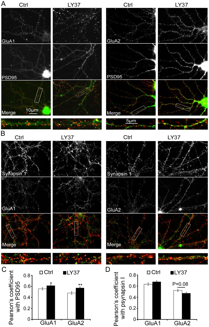
Group II metabotropic glutamate receptor (mGluR) agonists have emerged as potential treatment drugs for schizophrenia and other neurological disorders, whereas the mechanisms involved remain elusive. Here we examined the effects of LY379268 (LY37) on the expression and trafficking of the α-amino-3-hydroxy-5-methyl-4-isoxazole propionate (AMPA) receptor subunits GluA1 and GluA2 in prefrontal neurons. We show that LY37 significantly increased the surface and total expression of both GluA1 and GluA2 subunits in cultured prefrontal neurons and in vivo. This effect was mimicked by the selective mGluR2 agonist LY395756 and was blocked by mGluR2/3 antagonist LY341495. Moreover, we found that both GluA1 and GluA2 subunits were colocalized with PSD95 but not synapsin I, suggesting a postsynaptic localization. Consistently, treatment with LY37 significantly increased the amplitude, but not frequency, of miniature excitatory postsynaptic currents. Further, actinomycin-D blocked LY37's effects, suggesting a transcriptional regulation. In addition, application of glycogen synthase kinase-3beta (GSK-3β) inhibitor completely blocked LY37's effect on GluA2 surface expression, whereas GSK-3β inhibitor itself induced decreases in the surface and total protein levels of GluA1, but not GluA2 subunits. This suggests that GSK-3β differentially mediates GluA1 and GluA2 trafficking. Further, LY37 significantly increased the phosphorylation, but not total protein, of extracellular signal-regulated kinase 1/2 (ERK1/2). Neither ERK1/2 inhibitor PD98059 alone nor PD98059 combined with LY37 treatment induced changes in GluA1 or GluA2 surface expression or total protein levels. Our data thus suggest that mGluR2/3 agonist regulates postsynaptic AMPA receptors by affecting the synaptic trafficking of both GluA1 and GluA2 subunits and that the regulation is likely through ERK1/2 signaling in GluA1 and/or both ERK1/2 and GSK-3β signaling pathways in the GluA2 subunit.


Conflict of interest statement

Competing Interests: The authors have declared that no competing interests exist.

Figures

Figure 1
Figure 1. The mGluR2/3 agonist LY37 induces a significant increase in surface expression of GluA1 subunits in cultured PFC neurons.
(A) Representative immunofluorescent images of GluA1 receptor surface expression in response to a 60 min exposure to 0.1, 1, 10 or 100 µM LY37 treatment. LY37 at doses of 1, 10 and 100 µM increased the surface expression, i.e., cluster density, of GluA1 subunits on the dendrites. Images were taken by microscope at 63×oil lens. Scale bar = 10 µm for upper panels and for the enlarged images in the lower panels. (B) Pooled data show that surface puncta density of GluA1 was significantly increased by LY37 at doses of 1.0 to 100 µM (n = 20 neurons for control and for each doses, * p<0.05, ** p<0.01; ANOVA F = 3.501, p<0.01).
Figure 2
Figure 2. LY37 increased GluA2 surface expression in the cultured PFC neurons.
(A) Representative immunofluorescent images of GluA2 receptor surface expression in response to a 60 min exposure to 0.1, 1, 10, or 100 µM LY37 treatment. Culture medium served as a vehicle control. Scale bar = 10 µm for both upper panels and for the enlarged images in the lower panels. (B) Bar graphs show that GluA2 receptor surface puncta density on the neuronal dendrites was significantly increased by LY37 at concentrations from 1 to 100 µM (n = 20 neurons for each group, ** p<0.01). However, at lower doses of 0.01 and 0.1 µM, LY37 did not induce clear alterations in cluster numbers in GluA2 subunits (p > 0.05; ANOVA F = 5.288, p<0.001).
Figure 3
Figure 3. LY37 and selective mGluR2 agonist LY395756 (LY39) increased both surface and total protein level of GluA1 and GluA2 subunits in the cultured prefrontal neurons.
(A and B) Treatment with LY37 or LY395756 significantly increased the total protein levels of both GluA1 and GluA2 subunits (*p<0.05, **p<0.01, 6 independent experiments). (C) Representative immunoblots (left) show the surface and intracellular components of GluA1 and GluA2 subunits that were separated with BS3 cross-linker (right and middle panel). The surface proteins cross-linked with BS3 and formed large molecules at ∼500 KD, which were easily separated from the small intracellular components (∼100 KD) by Western Blot. In contrast, actin was detected only in 42KD because it is highly enriched in the cytosol but not in the cell membrane. Summary bar graphs on the right show the relative changes of surface and intracellular components of GluA1 and GluA2 subunits, respectively. LY37 (1 µM) treatment significantly increased the surface expression of both GluA1 and GluA2 (*p<0.05, 4 independent experiments). The intracellular and total protein levels of GluA2 subunit were also significantly increased by LY37 (*p<0.05, ** p<0.01). However, neither intracellular nor total protein level of GluA1 exhibited significance although there was a trend of increases (p > 0.05). (D) Presence of LY39 (1 µM) for 1 h in 17–18 DIV PFC neurons in incubator elevated the surface, intracellular, and total protein levels of GluA1 significantly; whereas the surface and total protein levels, but not the intracellular protein level, of GluA2 were significantly increased (*p<0.05, **p<0.01, 4 independent experiments).
Figure 4
Figure 4. Selective mGluR2/3 antagonist LY341495 (LY34, 100 nM) completely blocked the effects of LY37 and LY395756 (LY39) on the protein levels of both GluA1 and GluA2 subunits.
(A) LY37 signficantly increased the expression of GluA1 and GluA2 receptors, including surface, intracellular and total proteins (* p<0.05, ** p<0.01 compared with control). Further, whereas mGluR2/3 antagonist LY34 itself did not induce changes in protein levels of GluA1 or GluA2, LY34 completely blocked LY37's effects on protein levels of both GluA1 and GluA2 subunits when it was applied 30 min prior to LY37 treatment (p > 0.05 for all compared with control; # p<0.05, ## p<0.01 compared with LY37; 6 independent experiments). (B) Compared with control, LY39 signficantly increased the protein levels of surface, intracellular and total GluA1 and GluA2 receptors (* p<0.05, ** p<0.01) whereas LY34 itself did not induce changes in the protein levels of GluA1 and GluA2 receptors (p > 0.05 for all). However, LY34 effectively blocked LY39's effects on protein levels of both GluA1 and GluA2 (; # p<0.05, ## p<0.01 compared with LY39; 6 independent experiments).
Figure 5
Figure 5. LY37 increased GluA1 or GluA2 colocalization with PSD95 but not synapsin I.
(A) Immunofluorescent images show colocalization of surface GluA1 or GluA2 receptor (red: Dylight-594) with PSD95 (green: Alexa-594) in DIV15–17 primary PFC neurons. Scale bar = 10 µm for most of the images in A and B and scale bar = 5 µm in the enlarged images. (B) Coimmunolabeling of GluA1 (green: Alexa-488; control: n = 38 neurons, LY: n = 36), GluA2 (green: Alexa-488; control: n = 20, LY: n = 22) and synapsin I (red: Dylight-594) expression in cultured primary PFC neurons. (C) Bar graph shows the Pearson's correlation coefficients, in which LY37 (1 µM, 1 h) significantly increased the colocalization percentages of both GluA1 (control: n = 24, LY37: n = 31) and GluA2 surface receptors with PSD95 (control: n = 35, LY37: n = 39; *p< 0.05, **p<0.01). (D) Bar graph shows the percent colocalization of GluA1 or GluA2 with psynapsin I in neurons with or without LY37 treatment. The Pearson's correlation coefficient was similar, without significance for both GluA1 and GluA2 (p > 0.05 for both).
Figure 6
Figure 6. LY37 increased amplitude of AMPA receptor-mediated miniature excitatory postsynaptic currents (mEPSCs).
(A) Representative traces of mEPSCs were recorded from 15–18 DIV PFC neurons with (n = 12) or without (n = 10) treatment of LY37 (B) Bar graphs show that the amplitude of mEPSCs was significantly increased (*p<0.05; left) but the frequency of mEPSCs was unchanged (p > 0.05; Right) after treatment with LY37 (1 µM) for 1 hour at 37°C with culture medium.
Figure 7
Figure 7. Actinomycin-D partially blocks the effects of LY37 on total protein levels of GluA1 and GluA2 receptors.
(A) Representative blots of GluA1 and GluA2 in cultured PFC neurons in the presence or absence of Act-D (10 µg/ml, 4 h) prior to LY37 (1 µM, 1 h) treatment. (B) Compared with control, LY37 significantly increased total protein levels of GluA1 and GluA2 (* p<0.05 compared with control), whereas prior treatment with Actinomycin-D partially blocked these effects (p > 0.05 compared with control, 4 independent experiments).
Figure 8
Figure 8. Both GluA1 and GluA2 receptors, including surface, intracellular and total protein expressions, are significantly increased after the animals (P90 male SD rats) were treated with LY37 (0.3 mg/kg, i.p. injection, 1 h).
(A) Representative images show the GluA1 and GluA2 surface and intracellular receptors were separated by BS3 after gel electrophoresis. (B) Summary data show that LY37 in vivo injection significantly increased the expression levels in surface, intracellular, and total proteins of GluA1 and GluA2 (*p<0.05, **p<0.01; n = 6).
Figure 9
Figure 9. Specific GSK-3β inhibitor TDZD exhibited differential effects on the GluA1 and GluA2 subunit changes induced by LY37 treatment in cultured PFC neurons.
(A) Representative Western blot images show the surface and intracellular components of GluA1 and GluA2 subunits that were separated with BS3 cross-linker. (B and C) Summary bar graphs show the relative changes of surface and intracellular components of GluA1 and GluA2 subunits, respectively. It appears that TDZD itself significantly decreased the surface, intracellular, and total protein levels of GluA1 subunits but had no effects on the protein levels of GluA2 subunits (*p<0.05, ** p<0.01). However, when TDZD and LY were treated together simultaneously, the GluA1 expression, including surface, intracellular, and total protein levels, recovered to the control levels without significant differences (p > 0.05 for all). There were also no changes in GluA2 protein level when TDZD and LY were applied together (p > 0.05 for all). These data indicate that GSK-3β may not mediate the trafficking of GluA1 but is involved in the expression and trafficking of GluA2 receptor subunits.
Figure 10
Figure 10. LY37 treatment significantly increased the phosphorylation of ERK1/2 activity in the cultured PFC neurons, and this was prevented by the ERK inhibitor PD98059 (PD).
(A) Representative images of ERK1/2 and p-ERK1/2 expression in response to LY37, PD (50 µM for 1 h) or PD followed by LY37 treatment (1 µM for 1 h), DMSO as PD's vehicle. (B) LY37 administration did not induce clear changes in either ERK1 or ERK2 total protein level (p > 0.05). However, it significantly increased the expression of both p-ERK1 and p-ERK2 ratios (**p<0.01). PD significantly decreased both p-ERK1 and p-ERK2 protein ratios (*p <0.05, ** p <0.01) but not on the total protein levels of either ERK1 or ERK2 (P > 0.05 for both). With application of PD98059, LY37 did not affect the protein levels of p-ERK1 and p-ERK2 nor reverse the effects of PD98059.
Figure 11
Figure 11. ERK1/2 inhibitor PD98059 prevents the increases in surface levels of both GluA1 and GluA2 subunits induced by LY37 treatment in cultured PFC neurons.
(A) Representative images of drug treatment. Primary cultured PFC neurons at 17–18 DIV were exposed to DMSO (0.02%), LY37 (1 µM), ERK inhibitor PD98059 (50 µM), or PD98059 (50 µM) + LY37 (1 µM) for 1 h at 37°C, respectively. After treatments, the cells were incubated in BS3 cross-linker and the protein levels on the membrane surface and intracellular components were then separated by Western blot. (B and C) Summary bar graphs show the protein levels of surface and intracellular GluA1 and GluA2 subunits in different treatments groups compared to vehicle controls. Neither ERK1/2 inhibitor PD98059 nor PD98059+LY37 treatment induced changes in GluA1 and GluA2 surface expression (p > 0.05 for all comparisons).

Similar articles

Cited by

References

    1. Lyon L, Burnet PW, Kew JN, Corti C, Rawlins JN, et al. (2011) Fractionation of spatial memory in GRM2/3 (mGlu2/mGlu3) double knockout mice reveals a role for group II metabotropic glutamate receptors at the interface between arousal and gognition. Neuropsychopharmacology 36: 2616–2628. - PMC - PubMed
    1. Niswender CM, Conn PJ (2010) Metabotropic glutamate receptors: physiology, pharmacology, and disease. Annu Rev Pharmacol Toxicol 50: 295–322. - PMC - PubMed
    1. Fell MJ, McKinzie DL, Monn JA, Svensson KA (2012) Group II metabotropic glutamate receptor agonists and positive allosteric modulators as novel treatments for schizophrenia. Neuropharmacology 62: 1473–1483. - PubMed
    1. Conn PJ, Lindsley CW, Jones CK (2009) Activation of metabotropic glutamate receptors as a novel approach for the treatment of schizophrenia. Trends in Pharmacol Sci 30: 25–31. - PMC - PubMed
    1. Harrison PJ, Lyon L, Sartorius LJ, Burnet PW, Lane TA (2008) The group II metabotropic glutamate receptor 3 (mGluR3, mGlu3, GRM3): expression, function and involvement in schizophrenia. J Psychopharmacol 22: 308–322. - PubMed

Publication types

MeSH terms

Substances

LinkOut - more resources