. 2013 Apr 4;7(4):e2164.
doi: 10.1371/journal.pntd.0002164. Print 2013.

Combined TLR7/8 and TLR9 ligands potentiate the activity of a Schistosoma japonicum DNA vaccine

Affiliations

Combined TLR7/8 and TLR9 ligands potentiate the activity of a Schistosoma japonicum DNA vaccine

Xuefeng Wang et al. PLoS Negl Trop Dis. .

Abstract

Background: Toll-like receptor (TLR) ligands have been explored as vaccine adjuvants for tumor and virus immunotherapy, but few TLR ligands affecting schistosoma vaccines have been characterized. Previously, we developed a partially protective DNA vaccine encoding the 26-kDa glutathione S-transferase of Schistosoma japonicum (pVAX1-Sj26GST).

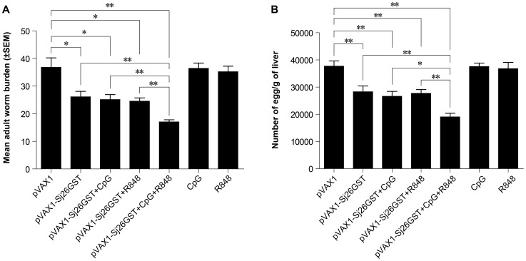
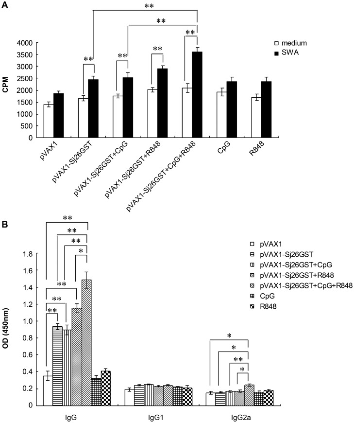
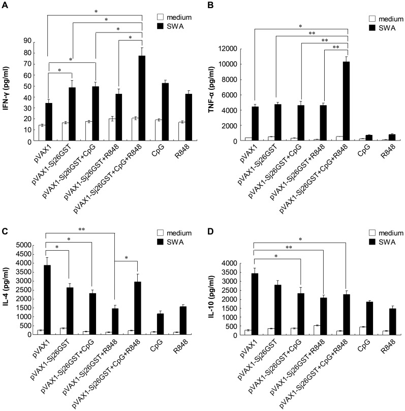
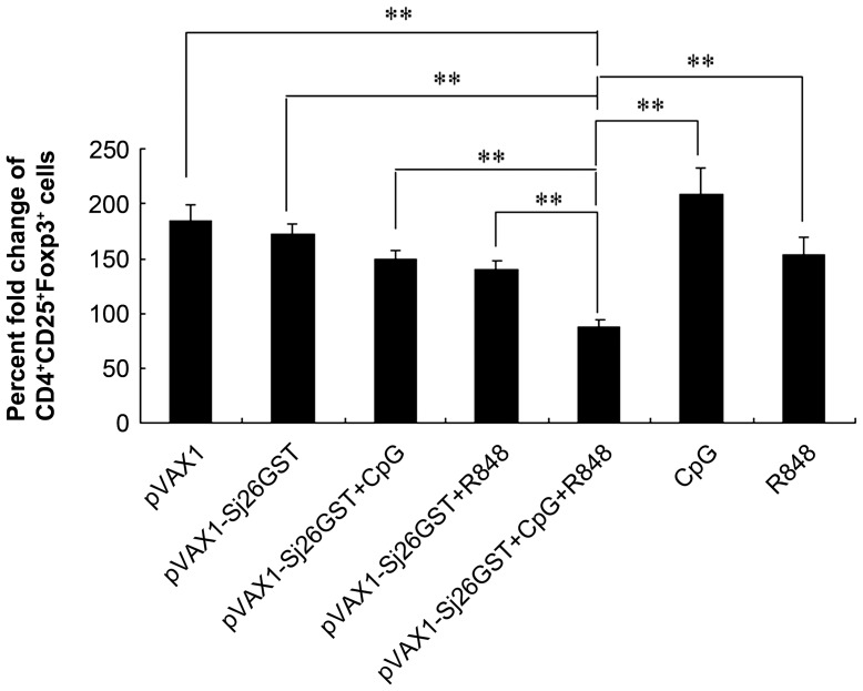
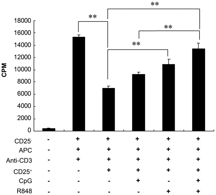
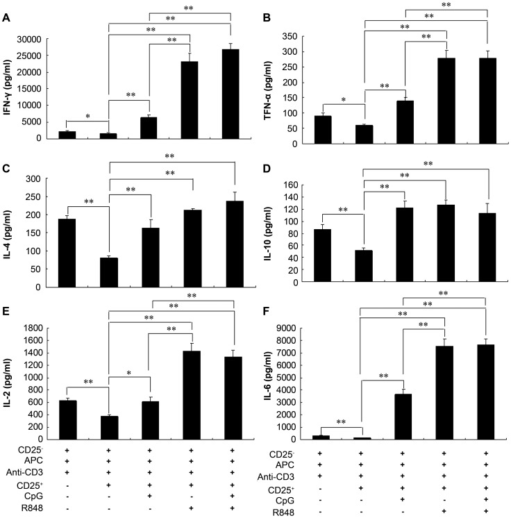
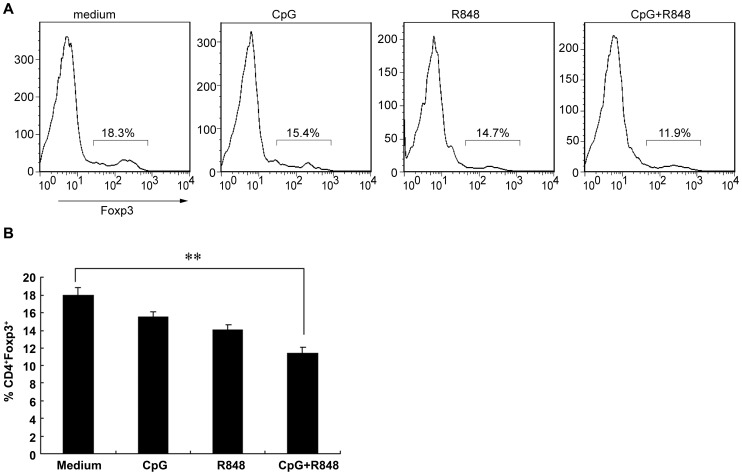
Methodology/principal findings: In this study, we evaluated a TLR7/8 ligand (R848) and a TLR9 ligand (CpG oligodeoxynucleotides, or CpG) as adjuvants for pVAX1-Sj26GST and assessed their effects on the immune system and protection against S. japonicum. We show that combining CpG and R848 with pVAX1-Sj26GST immunization significantly increases splenocyte proliferation and IgG and IgG2a levels, decreases CD4(+)CD25(+)Foxp3(+) regulatory T cells (Treg) frequency in vivo, and enhances protection against S. japonicum. CpG and R848 inhibited Treg-mediated immunosuppression, upregulated the production of interferon (IFN)-γ, tumor necrosis factor (TNF)-α, interleukin (IL)-4, IL-10, IL-2, and IL-6, and decreased Foxp3 expression in vitro, which may contribute to prevent Treg suppression and conversion during vaccination and allow expansion of antigen-specific T cells against pathogens.

Conclusions: Our data shows that selective TLR ligands can increase the protective efficacy of DNA vaccines against schistosomiasis, potentially through combined antagonism of Treg-mediated immunosuppression and conversion.


Conflict of interest statement

The authors have declared that no competing interests exist.

Figures

Figure 1
Figure 1. The combination of CpG and R848 increases protection induced by immunization with pVAX1-Sj26GST.
C57BL/6 mice (6 mice per group) were injected with pVAX1-Sj26GST (50 µg), with or without 25 µg CpG, 25 µg R848, or both CpG and R848 (CpG+R848). Seven days after the last dose of vaccine, mice were sacrificed for the characterization of cellular and humoral immune responses. Alternately, 2 weeks after the final vaccination, mice from each group (8 per group) were challenged percutaneously with 40±1 S. japonicum cercariae. Six weeks later, the mice were sacrificed and perfused to determine worm burdens and liver egg burdens. The adult worm (A) and liver egg burdens (B) per mouse in each group were determined. The data are expressed as the mean ± SEM (n = 8) and are representative of 2 independent experiments. * P<0.05; ** P<0.01.
Figure 2
Figure 2. The combination of CpG and R848 enhances splenocyte proliferation and antibody production in pVAX1-Sj26GST–vaccinated mice.
(A) Seven days after the last immunization with pVAX1-Sj26GST alone or with CpG, R848, or both (CpG+R848), splenocytes were harvested and antigen-specific proliferation was measured. Splenocytes (2×10⁵/well) from each mouse were incubated in the presence of SWA (15 µg/mL) or control media for 3 days in a 200 µL volume in 96-well plates. [3H] thymidine (0.5 µCi) was added to each well 16 h before the end of the incubation period. Data are expressed as the mean ± SEM (n = 6 per group) and are representative of 3 independent experiments performed in triplicate wells. * P<0.05. (B) IgG, IgG1, and IgG2a responses in immunized mice. Antibody responses to SWA (15 µg/mL) were determined by ELISA. Data are expressed as the mean ± SEM (n = 6 per group) of 18 mice from 3 independent experiments performed in triplicate wells. * P<0.05; ** P<0.01.
Figure 3
Figure 3. The combination of CpG and R848 upregulates proinflammatory cytokines in pVAX1-Sj26GST–vaccinated mice.
Seven days after the last immunization with pVAX1-Sj26GST, alone or with CpG, R848, or both (CpG+R848), splenocytes (2×10⁵/well) from each mouse were incubated in in the presence of SWA (15 µg/mL) or control media for 3 days in a 200-µL volume in 96-well plates. Supernatants were collected after 72 h and tested for IFN-γ (A), TNF-α (B), IL-4 (C), or IL-10 (D). Bars represent the mean ± SEM (n = 6 per group) of 18 mice from 3 independent experiments performed in triplicate wells. * P<0.05; ** P<0.01.
Figure 4
Figure 4. The combination of CpG and R848 inhibits the induction of CD4+CD25+ Tregs in pVAX1-Sj26GST–vaccinated mice.
Seven days after the last immunization with pVAX1-Sj26GST, alone or with CpG, R848, or both (CpG+R848), splenocytes from each mouse were analyzed by flow cytometry analysis for CD3, CD4, CD25, and Foxp3 expression. The mean fold change of the percentage of CD4+CD25+Foxp3+ Tregs in mice at week 0 compared with 5 weeks after immunization with the indicated formulations is shown. Groups consisted of 6 mice at each time point. The results are expressed as the mean ± SEM of 12 mice from 2 independent experiments. ** P<0.01.
Figure 5
Figure 5. The combination of CpG and R848 inhibits CD4+CD25+ Tregs function in vitro.
Isolated CD4+CD25 T cells (1×10⁵/well) from naïve mice were cultured with or without CD4+CD25+ Tregs (5×10⁴/well), irradiated APC (1×10⁵/well) and 1 µg/mL anti-CD3 in 96-well plates in the presence or absence of 3 µg/mL CpG, R848, or both for 72 h. Proliferation was determined by measuring 3H-thymidine incorporation for the last 16 h of the experiment. Data are expressed as the mean ± SEM of 3 experiments performed in triplicate. ** P<0.01.
Figure 6
Figure 6. R848 or its combination with CpG induces a panel of proinflammatory cytokines in a conventional in vitro suppression assay.
Isolated CD4+CD25 T cells (1×10⁵/well) from naïve mice were cultured with or without CD4+CD25+ Tregs (5×10⁴/well), irradiated APC (1×10⁵/well) and 1 µg/mL anti-CD3 in 96-well plates in the presence or absence of 3 µg/mL CpG, R848, or both. Supernatants were collected after 72 h and tested for IFN-γ (A), TNF-α (B), IL-4 (C), IL-10 (D), IL-2 (E), or IL-6 (F). Data are expressed as the mean ± SEM from 2 experiments performed in triplicate wells. * P<0.05; ** P<0.01.
Figure 7
Figure 7. The combination of CpG and R848 reduces the induction of Foxp3-expressing T cells in vitro.
Splenocytes from naïve mice were stimulated in vitro with 3 µg/mL CpG, R848, or both for 48 h and then subjected to flow cytometry analysis for CD3, CD4, CD25, and Foxp3 expression. (A) Representative flow cytometry data indicating the percentages of Foxp3 expression in CD4+ T cells. (B) The percentage of CD4+Foxp3+ T cells. Data are expressed as the mean ± SEM of 12 mice from 2 independent experiments. ** P<0.01.

References

    1. Hotez PJ, Brindley PJ, Bethony JM, King CH, Pearce EJ, et al. (2008) Helminth infections: the great neglected tropical diseases. J Clin Invest 118: 1311–1321. - PMC - PubMed
    1. King CH (2009) Toward the elimination of schistosomiasis. N Engl J Med 360: 106–109. - PubMed
    1. Doenhoff MJ, Cioli D, Utzinger J (2008) Praziquantel: mechanisms of action, resistance and new derivatives for schistosomiasis. Curr Opin Infect Dis 21: 659–667. - PubMed
    1. Abdul-Ghani R, Loutfy N, el-Sahn A, Hassan A (2009) Current chemotherapy arsenal for schistosomiasis mansoni: alternatives and challenges. Parasitol Res 104: 955–965. - PubMed
    1. Fenwick A, Webster JP, Bosque-Oliva E, Blair L, Fleming FM, et al. (2009) The Schistosomiasis Control Initiative (SCI): rationale, development and implementation from 2002–2008. Parasitology 136: 1719–1730. - PubMed

Publication types

MeSH terms