Review
. 2013 Apr 17;20(1):23.
doi: 10.1186/1423-0127-20-23.

Remodeling of calcium signaling in tumor progression

Affiliations
Review

Remodeling of calcium signaling in tumor progression

Yih-Fung Chen et al. J Biomed Sci. .

Abstract

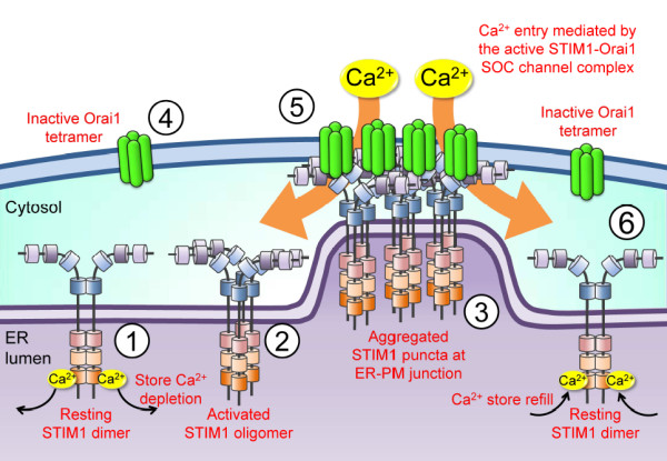
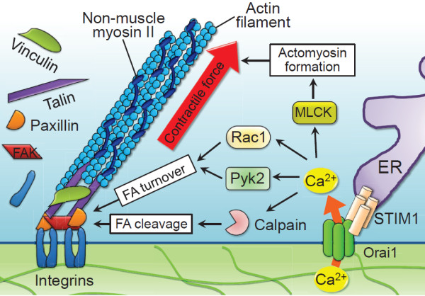
Intracellular Ca2+ is one of the crucial signalings that modulate various cellular functions. The dysregulation of Ca2+ homeostasis has been suggested as an important event in driving the expression of the malignant phenotypes, such as proliferation, migration, invasion, and metastasis. Cell migration is an early prerequisite for tumor metastasis that has a significant impact on patient prognosis. During cell migration, the exquisite spatial and temporal organization of intracellular Ca2+ provides a rapid and robust way for the selective activation of signaling components that play a central role in cytoskeletal reorganization, traction force generation, and focal adhesion dynamics. A number of known molecular components involved in Ca2+ influx pathways, including stromal interaction molecule (STIM)/Orai-mediated store-operated Ca2+ entry (SOCE) and the Ca2+-permeable transient receptor potential (TRP) channels, have been implicated in cancer cell migration and tumor metastasis. The clinical significance of these molecules, such as STIM proteins and the TRPM7 channel, in tumor progression and their diagnostic and prognostic potentials have also been demonstrated in specific cancer types. In this review, we summarize the recent advances in understanding the important roles and regulatory mechanisms of these Ca2+ influx pathways on malignant behaviors of tumor cells. The clinical implications in facilitating current diagnostic and therapeutic procedures are also discussed.


Figures

Figure 1
Figure 1
Regulatory mechanism of [Ca2+]i homeostasis. (A) Store-operated calcium entry (SOCE) in non-excitable cells. The activation of surface receptors stimulates phospholipase C (PLC) to increase the second messenger inositol-1,4,5-trisphosphate (IP3), which binds to the IP3 receptor in the endoplasmic reticulum (ER) membrane and causes rapid and transient Ca2+ release from the ER lumen. The decrease in ER luminal Ca2+ results in the opening of the plasmalemmal store-operated Ca2+ (SOC) channel, leading to the elevated intracellular Ca2+ levels ([Ca2+]i). (B) Domain architecture of the ER Ca2+ sensor stromal interaction molecule 1 (STIM1) protein. STIM1 is a single-transmembrane protein that is mainly localized in the endoplasmic reticulum (ER). The luminal N-terminus contains a canonical EF hand motif that binds Ca2+, a hidden EF hand that does not bind Ca2+, and a sterile α-motif (SAM) domain that is important for STIM1 oligomerization. The cytosolic C-terminus contains the coiled-coil domains, a STIM-Orai activating region (SOAR) or CRAC activation domain (CAD), and serine or proline (S/P)-rich segments and lysine (K)-rich clusters. The SOAR/CAD domain is essential for the gating of Orai1. The predicted protein-protein interaction domains in STIM1 include the SAM domain, coiled-coil domains, SOAR/CAD domain, S/P-rich segments and K-rich clusters. (C) Predicted topology of the plasmalemmal SOC channel Orai1. Orai1 consists of four transmembrane domains (TM1-TM4) and intracellular N- and C-termini. It is suggested that the TM1 lines the central pore of Orai1 channel. The short C-terminal putative coiled-coil domain is important for binding to the SOAR/CAD domain of STIM1, and its disruption impairs STIM1-mediated activation of Orai1 channel. Hexagonal structures represent glycosyl residues attached to an arginine (N)-linked glycosylation site (N223).
Figure 2
Figure 2
STIM1-dependent SOCE activation. STIM1 exists as the dimer maintained by the intermolecular interactions between its coiled-coil domains in the resting state (①). Depletion of ER luminal Ca2+ causes Ca2+ dissociation from the STIM1 N-terminal canonical EF hand, leading to STIM1 oligomerization due to the intermolecular interactions between EF-SAM domains (②). This activated STIM1 puncta interacts with the plasma membrane (PM) by the C-terminal polybasic K-rich clusters and accumulates in the ER-PM junctions (③). Orai1 tetramers diffusing in the PM (④) are tethered and trapped in junctions by the electrostatic interaction between CAD/SOAR domain in STIM1 and basic domains in the C-terminus of Orai1. The formation of the active STIM1/Orai1 complex conformationally gates the opening of SOC channel Orai1, thereby allowing Ca2+ entry (⑤). Upon store refilling, re-association of Ca2+ with STIM1 reverses EF-SAM oligomerization, causing STIM1-Orai1 uncoupling, Orai1 deactivation and the release of resting STIM1 dimers from puncta to redistribute throughout the ER (⑥).
Figure 3
Figure 3
STIM1-mediated Ca2+ influx regulates cell migration through focal adhesion turnover and actomyosin contractility. The dynamic interactions among cytoskeleton, non-muscle myosin II and cell-substrate adhesion regulate cell migration. The interaction between ER Ca2+ sensor STIM1 and plasma membrane SOC channel Orai1 induces Ca2+ influx, which integrates the dynamic interactions between actomyosin and focal adhesion (FA) to mediate efficient cell migration. STIM1/Orai1-mediated Ca2+ influx regulates actomyosin formation through the Ca2+-dependent myosin light chain kinase (MLCK), accelerates FA turnover through the small GTPase Rac1 and the Ca2+-dependent proline-rich tyrosine kinase 2 (Pyk2), and promotes calpain-mediated disassembly and cleavage of FA proteins.
Figure 4
Figure 4
STIM1/Orai1-mediated Ca2+ signalings in tumor biology. Ca2+ homeostasis is remodeled in cancer cells during tumor progression, with Ca2+ influx increasing through STIM1/Orai1upregulation. STIM1/Orai1-dependent Ca2+ signaling integrates the dynamic interactions between actomyosin contraction and focal adhesion turnover to mediate efficient cell migration. STIM1 also influences cancer cell proliferation through cell cycle regulators p21 and cdc25C. Additionally, STIM1-mediated SOCE plays an important role in tumor angiogenesis; one possible mechanism involves the production of vascular endothelial growth factor (VEGF) from cancer cells, another is the cell cycle progression of vascular endothelial cells [72]. Accordingly, STIM1/Orai1-remodeled Ca2+ homeostasis is important for aggravating tumor development in vivo.

References

    1. Berridge MJ, Bootman MD, Roderick HL. Calcium signalling: dynamics, homeostasis and remodelling. Nat Rev Mol Cell Biol. 2003;4:517–529. doi: 10.1038/nrm1155. - DOI - PubMed
    1. Carafoli E. The calcium-signalling saga: tap water and protein crystals. Nat Rev Mol Cell Biol. 2003;4:326–332. doi: 10.1038/nrm1073. - DOI - PubMed
    1. Rizzuto R, Pozzan T. When calcium goes wrong: genetic alterations of a ubiquitous signaling route. Nat Genet. 2003;34:135–141. doi: 10.1038/ng0603-135. - DOI - PubMed
    1. Parekh AB. Store-operated CRAC channels: function in health and disease. Nat Rev Drug Discov. 2010;9:399–410. doi: 10.1038/nrd3136. - DOI - PubMed
    1. Monteith GR, Davis FM, Roberts-Thomson SJ. Calcium channels and pumps in cancer: changes and consequences. J Biol Chem. 2012;287:31666–31673. doi: 10.1074/jbc.R112.343061. - DOI - PMC - PubMed

Publication types

LinkOut - more resources