Review
. 2013 May;13(5):342-55.
doi: 10.1038/nrc3495. Epub 2013 Apr 18.

Iron and cancer: more ore to be mined

Affiliations
Review

Iron and cancer: more ore to be mined

Suzy V Torti et al. Nat Rev Cancer. 2013 May.

Abstract

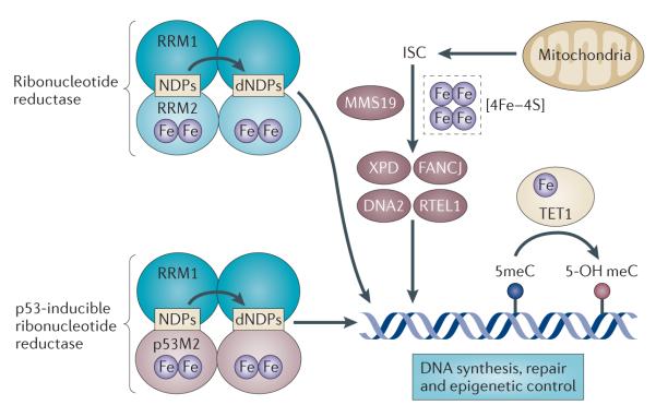
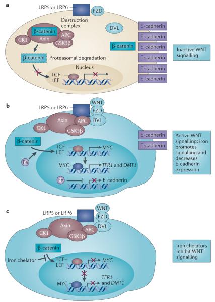
Iron is an essential nutrient that facilitates cell proliferation and growth. However, iron also has the capacity to engage in redox cycling and free radical formation. Therefore, iron can contribute to both tumour initiation and tumour growth; recent work has also shown that iron has a role in the tumour microenvironment and in metastasis. Pathways of iron acquisition, efflux, storage and regulation are all perturbed in cancer, suggesting that reprogramming of iron metabolism is a central aspect of tumour cell survival. Signalling through hypoxia-inducible factor (HIF) and WNT pathways may contribute to altered iron metabolism in cancer. Targeting iron metabolic pathways may provide new tools for cancer prognosis and therapy.


Figures

Figure 1
Figure 1. Key features of systemic iron homeostasis in humans
Dietary iron (predominantly in the form of ferric iron (Fe3+)) is absorbed in the duodenum through the concerted action of a reductase, such as duodenal cytochrome b (DCYTB), which produces ferrous iron (Fe2+), and divalent metal transporter 1 (DMT1). Iron exits the basolateral surface of the enterocyte through the iron efflux pump ferroportin, which functions together with the oxidase hephaestin to oxidize ferrous iron to form ferric iron, which is loaded onto transferrin (TF). The diferric iron transferrin complex (TF–[Fe3+]2) circulates through the bloodstream to deliver iron to sites of utilization. Principal among these sites is the bone marrow, where iron is used in the synthesis of haemoglobin and red blood cells (RBCs). RBCs circulate for approximately 90 days before they are catabolized by macrophages of the reticuloendothelial (RE) system. Iron is released from catabolized haem and effluxed out of the macrophage through the action of ferroportin, where it is loaded onto TF in the bloodstream, in a process termed iron recycling. TF–[Fe3+]2 is also delivered to peripheral tissues and the liver, which is the primary organ for the storage of excess iron. Although small amounts of iron are lost through desquamation, there is no excretory pathway for iron, so levels of iron in the body are primarily regulated at the absorption step. Excess iron induces the synthesis of the peptide hormone hepcidin (HP), which serves as a master regulator of systemic iron homeostasis. HP binds to ferroportin and triggers its degradation, inhibiting both delivery of dietary iron through the enterocyte and iron recycling through the macrophage. HP is also induced in response to inflammatory cytokines and thus contributes to the anaemia of cancer.
Figure 2
Figure 2. Key steps in mammalian cellular iron metabolism
Iron circulates throughout the body bound to transferrin (TF), which can bind two atoms of ferric iron (Fe3+). TF-bound iron binds to transferrin receptor 1 (TFR1) on the plasma membrane of most cells, and the TF– [Fe3+]2–TFR1 complex is endocytosed. In the acidic environment of the endosome, ferric iron is released from TF and is reduced to ferrous iron (Fe2+) through the ferrireductase activity of STEAP3. The apotransferrin–TFR1 complex then recycles back to the cell surface, where apotransferrin participates in further rounds of iron uptake. In the meantime, ferrous iron is transported out of the endosome into the cytosol by divalent metal transporter 1 (DMT1), and enters the metabolically active pool of iron (the labile iron pool). Iron then traffics to multiple destinations. It is inserted into cytosolic enzymes that are required for DNA synthesis, such as ribonucleotide reductase, and is also used in haem synthesis and the biogenesis of iron–sulphur clusters, processes that occur partly in the mitochondria and partly in the cytosol. Excess iron is stored in ferritin, an iron storage protein. Iron leaves the cell through the activity of ferroportin, an iron efflux pump, and an oxidase such as ceruloplasmin or hephaestin, which can re-oxidize iron to ferric iron to enable the loading onto TF.
Figure 3
Figure 3. Iron uptake and efflux in malignant and non-malignant cells
a | Normal epithelial cells express low levels of transferrin (TF) receptor 1 (TFR1) and hepcidin and high levels of ferroportin, which collectively lead to a small pool of labile iron. In breast cells, lipocalin 2 (LCN2), in a complex with a siderophore (SD), may further reduce levels of intracellular iron by capturing and effluxing SD-bound iron from these cells, although this is currently hypothetical. b | Cancer cells show increased expression of TFR1 and hepcidin and low levels of ferroportin, which lead to an increased labile iron pool. In breast cancer cells, LCN2, in a complex with SD-bound iron, may serve as a further source of iron. LCN2R, LCN2 receptor.
Figure 4
Figure 4. Control of cellular iron metabolism by the IRE–IRP regulatory axis
Iron-regulatory protein 1 (IRP1) and IRP2 are crucial proteins in the maintenance of cellular iron homeostasis. These proteins bind to iron-response elements (IREs) present in either the 5′ or the 3′ untranslated region (UTR) of mRNAs. IREs are found in the 5′ UTR of mRNAs encoding the ferritin heavy chain (FTH) and ferritin light chain (FTL) subunits, ferroportin and hypoxia-inducible factor 2α (HIF2α), and in the 3′ UTR of mRNAs encoding transferrin receptor 1 (TFR1) and IRE-containing isoforms of divalent metal transporter 1 (DMT1). Binding of IRPs to 5′ IREs inhibits translation, whereas binding to 3′ IREs stabilizes mRNA. IRPs bind to IREs under conditions of low iron levels; under conditions of high iron levels, IRP1 loses its IRE-binding activity and acquires enzymatic activity as a cytosolic aconitase, whereas IRP2 is degraded. Thus, under conditions of low iron levels, the IRE–IRP system functions to increase iron uptake (by stabilizing mRNAs that encode TFR1 and presumably IRE-containing isoforms of DMT1) and decreases iron storage and efflux (by inhibiting the translation of ferritin and ferroportin). Binding of IRPs to mRNAs encoding HIF2α may function as a feedback loop to inhibit erythropoiesis when iron levels are low.
Figure 5
Figure 5. Links between iron, DNA metabolism and genomic integrity
Iron is essential for the activity of the enzymes involved in DNA synthesis, DNA repair and epigenetic regulation. A di-iron site is essential for the catalytic activity of both constitutive and p53-inducible ribonucleotide reductase, the enzyme that catalyses the reductive conversion of ribonucleotides (NDPs) to deoxyribonucleotides (dNDPs) for DNA synthesis. MMS19 serves as a scaffold for the insertion of iron–sulphur clusters (ISCs) into DNA repair enzymes such as Xeroderma pigmentosum group D-complementing protein (XPD), Fanconi anaemia group J protein (FANCJ), DNA replication helicase 2 homologue (DNA2) and regulator of telomere elongation helicase 1 (RTEL1). The ferrous iron- and 2-oxoglutarate-dependent enzyme TET1 catalyses the hydroxylation of methylcytosines (meCs) in the DNA and may have a role in epigenetic control. RRM, ribonucleoside-diphosphate reductase subunit M.
Figure 6
Figure 6. A role for iron in canonical WNT signalling
a | In the absence of a trigger for WNT signalling, β-catenin associates with axin, adenomatous polyposis coli (APC), casein kinase 1 (CK1) and glycogen synthase kinase 3β (GSK3β) (collectively known as the destruction complex) and is targeted for degradation (shown by dashed outline). Simultaneously, β-catenin is sequestered from the destruction complex and associates with E-cadherin. b | In cells with constitutive canonical WNT signalling (such as cells with mutant APC), β-catenin evades destruction, enters the nucleus and promotes T cell factor (TCF)–lymphoid enhancer factor (LEF)-dependent transcription of downstream target genes such as MYC. Iron promotes TCF–LEF-dependent transcription in cells with such constitutive WNT signalling, resulting in the induction of MYC expression. MYC in turn transcriptionally induces transferrin receptor 1 (TFR1) and divalent metal transporter 1 (DMT1) to promote iron uptake. Iron also decreases E-cadherin mRNA and protein levels in an APC-independent manner. c | Iron chelators decrease TCF–LEF signalling in cells with constitutive WNT signalling at a step distal to β-catenin by an unknown mechanism. DVL, disheveled homologue; FZD, frizzled homologue; LRP, low-density lipoprotein receptor-related protein.

References

    1. Crichton R. Iron Metabolism: from Molecular Mechanisms to Cinical Consequences. John Wiley and Sons; 2009. pp. 17–58.
    1. Inoue S, Kawanishi S. Hydroxyl radical production and human DNA damage induced by ferric nitrilotriacetate and hydrogen peroxide. Cancer Res. 1987;47:6522–6527. - PubMed
    1. Dizdaroglu M, Rao G, Halliwell B, Gajewski E. Damage to the DNA bases in mammalian chromatin by hydrogen peroxide in the presence of ferric and cupric ions. Arch. Biochem. Biophys. 1991;285:317–324. - PubMed
    1. Dizdaroglu M, Jaruga P. Mechanisms of free radical-induced damage to DNA. Free Radic. Res. 2012;46:382–419. - PubMed
    1. Campbell JA. Effects of precipitated silica and of iron oxide on the incidence of primary lung tumours in mice. Br. Med. J. 1940;2:275–280. - PMC - PubMed

Publication types