. 2013 Jun;10(6):1003-8.
doi: 10.4161/rna.24454. Epub 2013 Apr 1.

Long non-coding RNAs as targets for cytosine methylation

Affiliations

Long non-coding RNAs as targets for cytosine methylation

Thomas Amort et al. RNA Biol. 2013 Jun.

Abstract

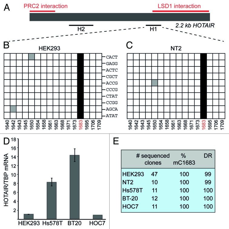
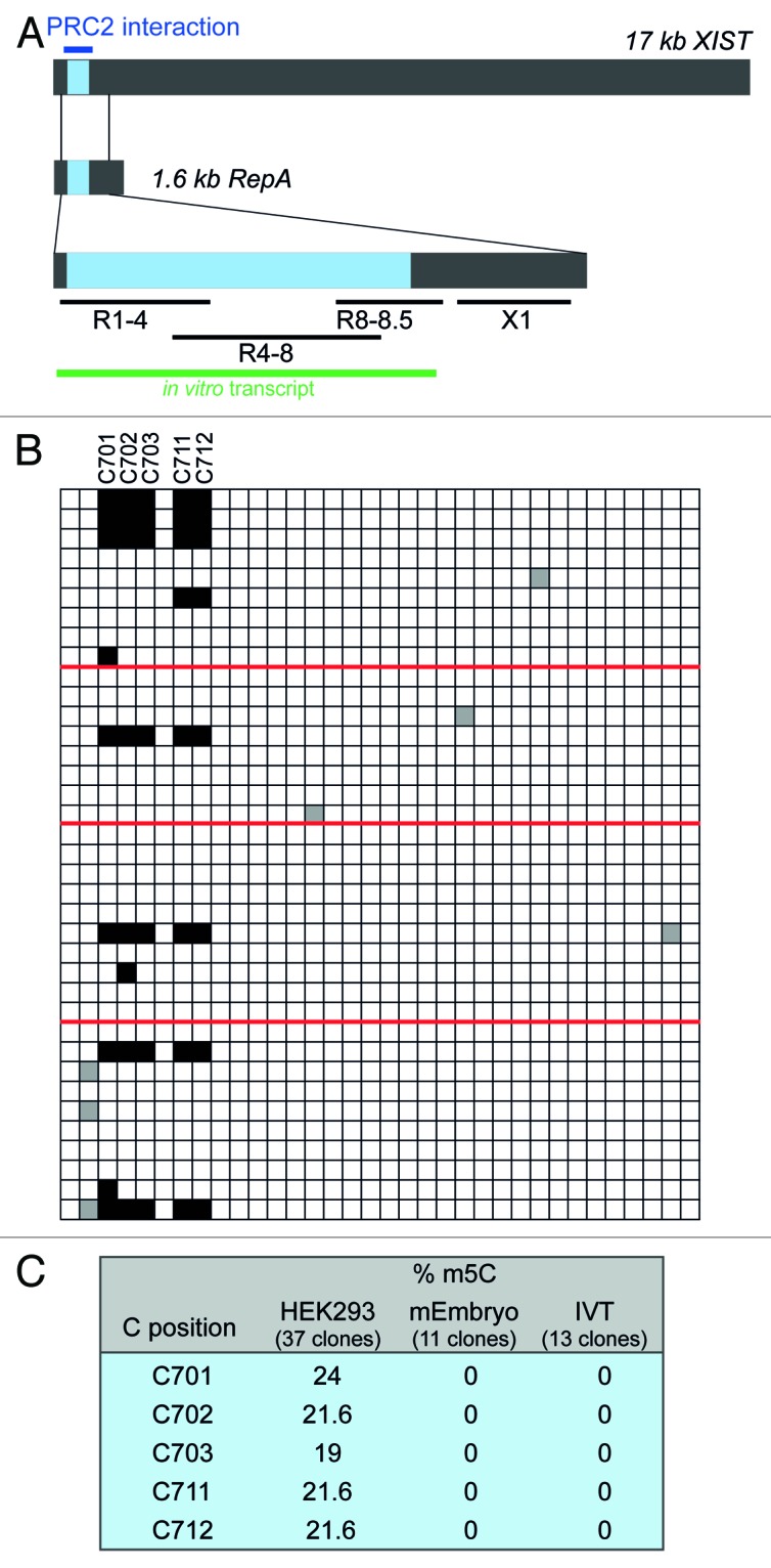
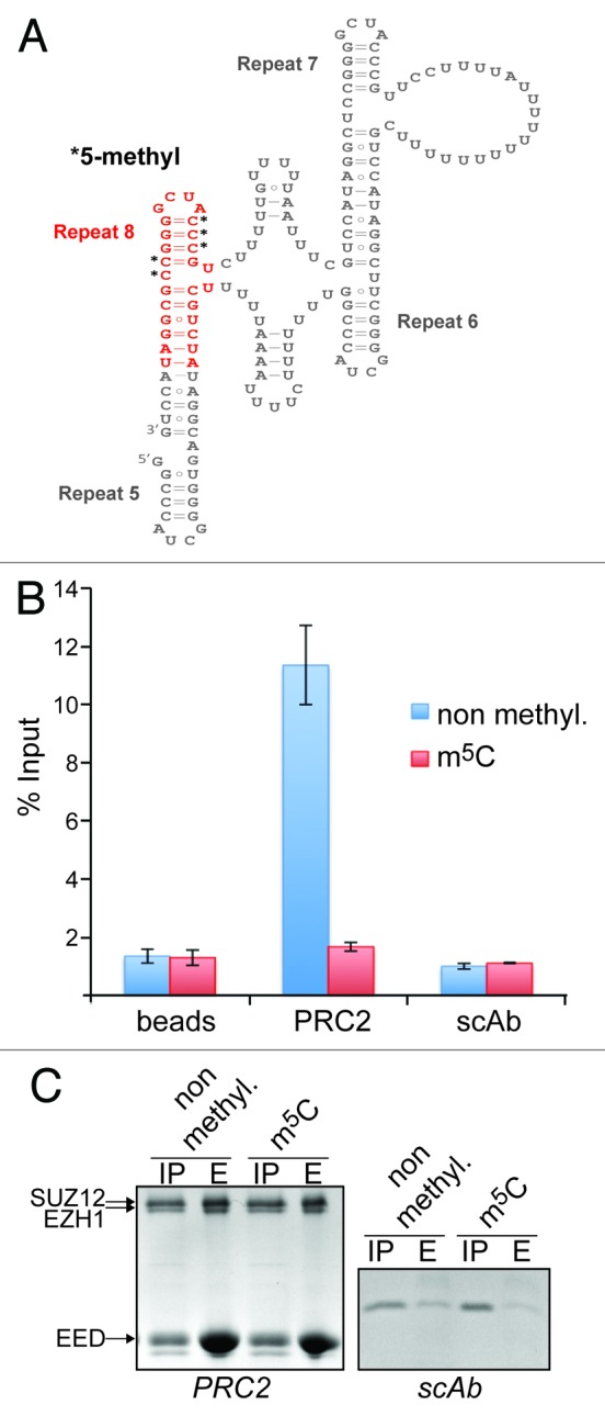
Post-synthetic modifications of nucleic acids have long been known to affect their functional and structural properties. For instance, numerous different chemical modifications modulate the structural organization, stability or translation efficiency of tRNAs and rRNAs. In contrast, little is known about modifications of poly(A)RNAs. Here, we demonstrate for the first time that the two well-studied regulatory long non-coding RNAs HOTAIR and XIST are targets of site-specific cytosine methylation. In both XIST and HOTAIR, we found methylated cytosines located within or near functionally important regions that are known to mediate interaction with chromatin-associated protein complexes. We show that cytosine methylation in the XIST A structure strongly affects binding to the chromatin-modifying complex PRC2 in vitro. These results suggest that cytosine methylation may serve as a general strategy to regulate the function of long non-coding RNAs.

Keywords: 5-methylcytosine; HOTAIR RNA; RNA methylation; XIST RNA; chromatin; long non-coding RNA; polycomb repressive complex 2.


Figures

None
Figure 1. HOTAIR lncRNA shows m5C in various cell types. (A) Schematic representation of HOTAIR RNA. Previously mapped PRC2 and LSD1-interacting regions are marked in red. Black lines designate the regions that were analyzed by BS sequencing. (B and C) Sequencing results of 10 clones derived from BS treated HOTAIR RNA from HEK293 (B) and NT2 (C) cells. Each horizontal row in the diagrams corresponds to one sequenced clone, each square to a specific C (numbers at the bottom indicate the position of the C within the HOTAIR sequence). Non-deaminated (i.e. methylated) Cs are shown as black squares, white squares are deaminated Cs and gray squares depict non-deaminated Cs that were considered BS treatment artifacts. Barcode labels of individual clones are indicated in (B). (D) Expression analysis of HOTAIR in the breast cancer cell lines Hs578T, BT20 and in HOC7 ovarian cancer cells. RT-qPCR results are expressed relative to values obtained from HEK293 cells. (E) Summary of BS sequencing results for HOTAIR in different cell lines; DR, deamination rate in percent.
None
Figure 2. Cytosine methylation within A-region repeat 8 of XIST. (A) Schematic representation of XIST RNA and the overlapping RepA transcript. The A region is shown in blue. Blue line, PRC2 interaction domain; black lines, regions analyzed by BS sequencing; green line, size and position of the A-region in vitro transcript. (B) BS sequencing results for region R8-8.5. Red lines demarcate results obtained from different experiments with fresh batches of cells. (C) Summary of BS sequencing results of R8-8.5 in different cell types as well as in in vitro transcribed (IVT) XIST templates.
None
Figure 3. Cytidine methylation in XIST repeat 8 prevents binding of the PRC2 complex. (A) Two-dimensional structure model of R5-R8 showing the position and sequence of the RNA used in the RNA-binding assay (marked in red). Black asterisks indicate the methylated cytidines. (B) Bar graph showing the quantification by scintillation counting of 32P-5′ end-labeled XIST R8 RNA bound to affinity-purified PRC2. Flag-tagged PRC2 or a Flag-tagged control protein (scAb) was incubated with either the unmodified RNA (blue bars) or RNA methylated on the 5 cytidines indicated in (A) (red bars) and subsequently purified by Flag-M2-agarose. In addition, the RNAs were incubated with anti-Flag-beads in the absence of protein (beads). Values represent mean +/− SD of three independent experiments. (C) Coomassie blue stained SDS-polyacrylamide gels showing input material (IP) and eluates (E) of Flag-M2-beads from reactions described in (B) for PRC2 (left panel) and scAb (right panel). Twenty-five percent and 100%, respectively, of input of PRC2 and scAb were loaded in the IP lanes, 30% of eluates were loaded in the E lanes. The positions of the PRC2 subunits EZH1 (86 kDa), SUZ12 (87 kDa) and EED (51 kDa) are indicated. The MW of scAb is 18 kDa.

References

    1. Hotchkiss RD. The quantitative separation of purines, pyrimidines, and nucleosides by paper chromatography. J Biol Chem. 1948;175:315–32. - PubMed
    1. Wyatt GR. Occurrence of 5-methylcytosine in nucleic acids. Nature. 1950;166:237–8. doi: 10.1038/166237b0. - DOI - PubMed
    1. Grosjean H. Nucleic acids are not boring long polymers of only four types of nucleotides: a guided tour. In: Grosjean H, ed. DNA and RNA Modification Enzymes: Structure, Mechanism, Function and Evolution. Austin, Texas: Landes Bioscience, 2009:1-18.
    1. Szyf M. Epigenetics, DNA methylation, and chromatin modifying drugs. Annu Rev Pharmacol Toxicol. 2009;49:243–63. doi: 10.1146/annurev-pharmtox-061008-103102. - DOI - PubMed
    1. Motorin Y, Helm M. RNA nucleotide methylation. Wiley Interdiscip Rev RNA. 2011;2:611–31. doi: 10.1002/wrna.79. - DOI - PubMed

Publication types

LinkOut - more resources