. 2013 Jul;126(1):59-74.
doi: 10.1007/s00401-013-1119-4. Epub 2013 Apr 18.

Mild traumatic brain injury in the mouse induces axotomy primarily within the axon initial segment

Affiliations

Mild traumatic brain injury in the mouse induces axotomy primarily within the axon initial segment

John E Greer et al. Acta Neuropathol. 2013 Jul.

Abstract

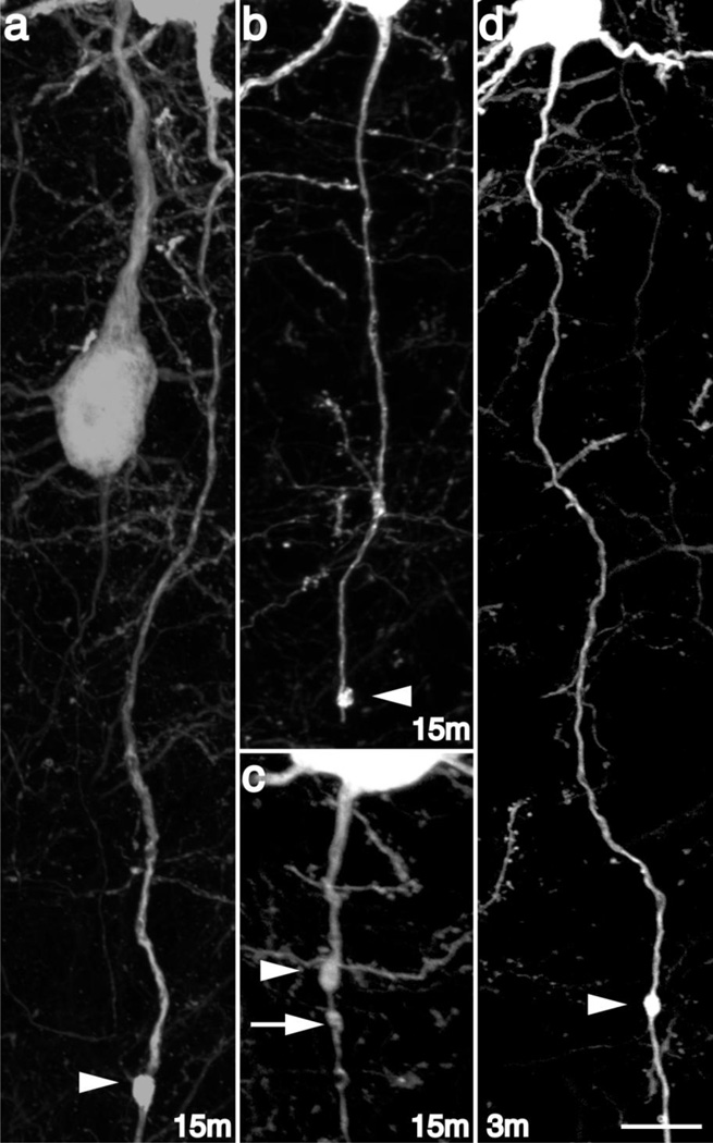
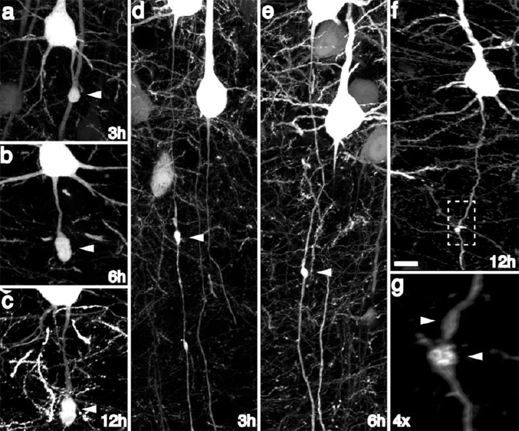
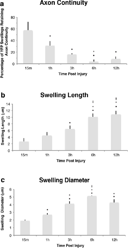
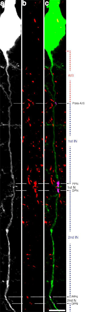
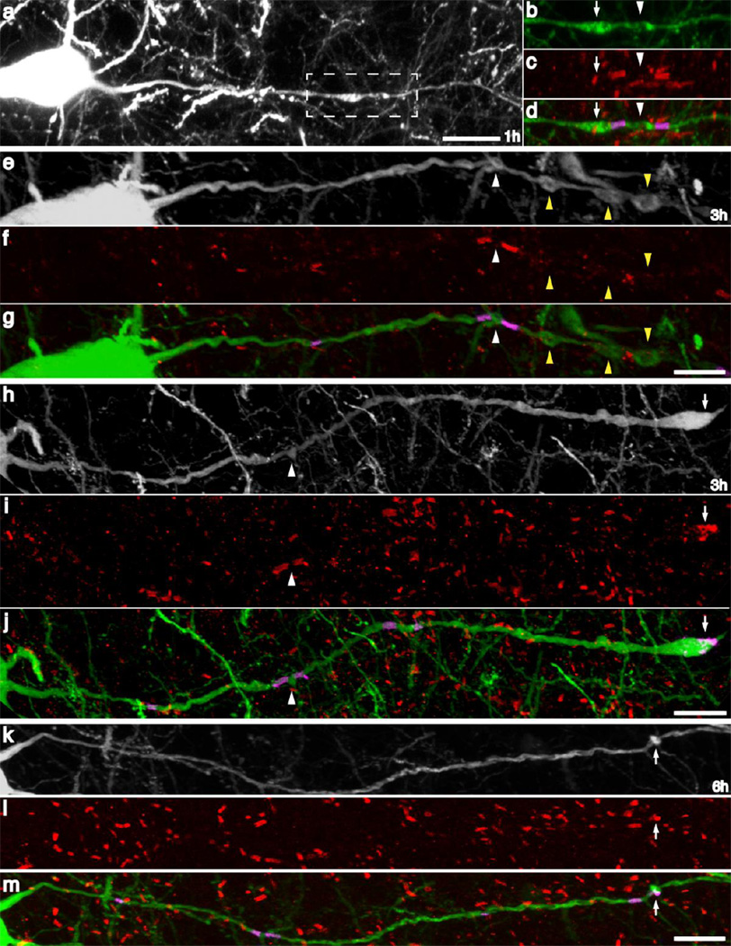
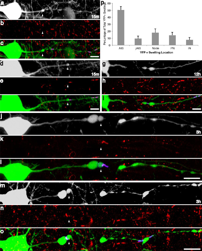
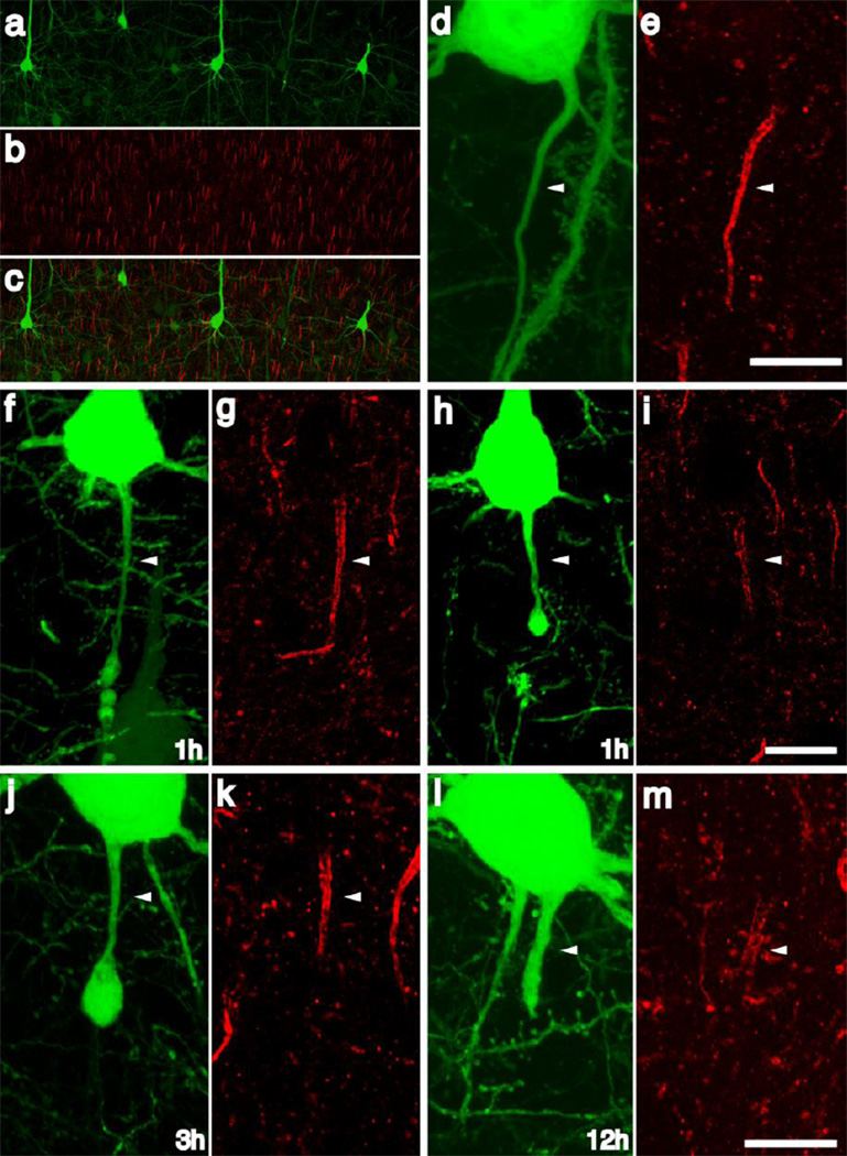
Traumatic axonal injury (TAI) is a consistent component of traumatic brain injury (TBI), and is associated with much of its morbidity. Increasingly, it has also been recognized as a major pathology of mild TBI (mTBI). In terms of its pathogenesis, numerous studies have investigated the susceptibility of the nodes of Ranvier, the paranode and internodal regions to TAI. The nodes of Ranvier, with their unique composition and concentration of ion channels, have been suggested as the primary site of injury, initiating a cascade of abnormalities in the related paranodal and internodal domains that lead to local axonal swellings and detachment. No investigation, however, has determined the effect of TAI upon the axon initial segment (AIS), a segment critical to regulating polarity and excitability. The current study sought to identify the susceptibility of these different axon domains to TAI within the neocortex, where each axonal domain could be simultaneously assessed. Utilizing a mouse model of mTBI, a temporal and spatial heterogeneity of axonal injury was found within the neocortical gray matter. Although axonal swellings were found in all domains along myelinated neocortical axons, the majority of TAI occurred within the AIS, which progressed without overt structural disruption of the AIS itself. The finding of primary AIS involvement has important implications regarding neuronal polarity and the fate of axotomized processes, while also raising therapeutic implications, as the mechanisms underlying such axonal injury in the AIS may be distinct from those described for nodal/paranodal injury.


Conflict of interest statement

Conflict of Interest

The authors declare that they have no conflict of interest.

Figures

Fig 1
Fig 1. Diffuse brain injury induces rapid secondary axotomy within Layer V pyramidal neurons
Following cFPI, the YFP+ axonal swellings (arrowheads) were evident as early as 15m post injury (a–c). Several injured YFP+ fibers at this time point following injury showed clear evidence for disconnection (b), though many demonstrated retained axonal continuity (a, c). Additionally, YFP+ swellings could be found at various sites along the YFP+ axon, both distal (a, b) and proximal (c) relative to the cell body of origin. Note the presence of multiple swellings along the length of some injured YFP+ fibers at this early time point (C, arrow/arrowheads). Importantly, axonal disconnection (b) at 15m following injury was the result of secondary and progressive axonal change, as limited axonal alterations were observed at 3–4m following injury, and the only YFP+ swelling noted remained in continuity with the distal axonal segment (d). Scale: 10 µm
Fig 2
Fig 2. YFP+ axonal swellings progressively enlarge and disconnect over time post-injury
Consistent with progressive axonal change, YFP+ swellings continue to enlarge over the next 12h post-injury (a–c). Though the majority of swellings progress to disconnection over this 12h time span, occasional swellings still maintaining axonal continuity could be found at later time points following injury (d–g), underscoring a temporally heterogeneous evolution of YFP+ swellings over time. Scale: 10 µm
Fig 3
Fig 3. Quantitative assessment of the temporal alteration in YFP+ swelling characteristics revealed rapid axonal disconnection following TAI
When quantified, a large number of injured axons demonstrated axonal disconnection at 15m post-injury (a). Reflecting progressive disconnection, the number of YFP+ axons demonstrating axonal continuity decreased significantly at 1h, 3h and 6h when compared to 15m post-injury, with most fibers demonstrating disconnection by 6h post-injury (a). Quantitative analysis of YFP+ swellings revealed significant increases in YFP+ swelling length (b) and diameter (c), reflecting progressive swelling enlargement over time. Importantly, (* p < 0.05, compared to 15m; + p < 0.05, compared to 1h; ± p < 0.05, compared to 3h)
Fig 4
Fig 4. β-Amyloid precursor protein accumulation marks the site of eventual axon disconnection
At 15m following injury APP+/YFP+ swellings could be identified within the neocortex (a, b, e, f). Within axons maintaining continuity, APP was consistently observed to be present in the more proximal swellings (e, f arrowheads) and typically excluded from more distal swellings (e, f arrows). Additional APP accumulation occurred within the larger YFP+ swellings at later time points (c, d). Importantly, as axons disconnected at later time points (f, g) APP localization remained consistent with that observed at 15m. Note the disconnected YFP+ axon, demonstrating APP immunoreactivity within the proximal YFP+ swelling (f, g arrowhead) with a lack of similar immunoreactivity within distal swellings (f, g arrows). Importantly, at later time points (i–j) were multiple swellings were found within disconnected axons, APP immunoreactivity was restricted to the swelling adjacent to the site of disconnection, suggesting that the extent of APP accumulation along the length of an injured YFP+ axon demarcates the region that will be retained following eventual disconnection. Scale: 10 µm
Fig 5
Fig 5. Caspr immunoreactivity demarcates multiple axon domains
Caspr immunoreactive bands located along YFP+ axons mark sites of axoglial interaction. Note the consistent pattern of immunoreactivity, with a proximal single band of Caspr immunoreactivity localized to the para-AIS and the beginning of myelination, delineating the more proximal AIS. More distal Caspr immunoreactivity appears with Caspr bands occurring in pairs, marking two adjacent paranodes, flanking the node of Ranvier. Scale: 10 µm
Fig 6
Fig 6. TAI occurs in all axon domains following cFPI
Utilizing Caspr immunoreactivity, YFP+ swellings following injury were often localized to paranodal and nodal regions (a–d, h–j). Less frequently, swellings were observed within the intermodal region between two Caspr+ nodal regions (e–g). At later time points, YFP+ swellings, lacking continuity, were often observed to have maintained a Caspr+ region within the distal region of the YFP+ swelling (h–j), presumably indicating disconnection within the distal nodal region and retention of one Caspr+ hemi node within the distal YFP+ swelling. Additionally, paranodal YFP+ swellings were also observed to occur without apparent morphological alteration of the adjacent nodal or internodal regions (k–m). Scale: 10 µm
Fig 7
Fig 7. Axonal injury following cFPI preferentially occurs in the axon initial segment
Following quantitative analysis, the majority of YFP+ axonal swellings were found to occur within the AIS (p). At 15m YFP+ swellings were predominantly localized at the end of the AIS, proximal to the para-AIS (d–f), though several swellings were found to occur within the AIS (a–c). At later time points and following disconnection, swellings were observed within the AIS (white arrowheads, j–1), injured YFP+ axons were often found to have retained Caspr immunoreactivity within the distal region of the swelling (j–o) or this Caspr+ axoglial interaction was clearly visible within the distal, disconnected YFP axonal segment (G–I). Scale: 10 µm
Fig 8
Fig 8. The AIS cytoskeletal integrity is conserved following traumatic axonal injury within the AIS
AnkG labeled the axon initial segments throughout the neocortex in sham-injured animals (a–c). Within intact axons of sham-injured and injured animals demonstrating no focal swellings, AnkG immunolabeling revealed a discrete and compact AIS associated with each YFP+ neuron (d, e). Within most YFP+ axons demonstrating swellings within the AIS, the AnkG cytoskeleton was conserved and consistent with the pattern observed within sham animals with loss of AnkG expression restricted to the region of axonal swelling (1h f, G; 3h j, k). Occasional axotomized neurons demonstrated attenuation of the AnkG expression within the AIS (h, i) and within YFP+ axons demonstrating expansion of the swelling to include the entire AIS ankG expression is still present directly underneath the plasmolemma (l, m). Scale: 10 µm

References

    1. Adams JH, Doyle D, Ford I, Gennarelli TA, Graham DI, McLellan DR. Diffuse axonal injury in head injury: definition, diagnosis and grading. Histopathology. 1989;15(1):49–59. - PubMed
    1. Baalman KL, Cotton RJ, Rasband SN, Rasband MN. Blast wave exposure impairs memory and decreases axon initial segment length. J Neurotrauma. 2013 - PMC - PubMed
    1. Banik NL, Matzelle DC, Gantt-Wilford G, Osborne A, Hogan EL. Increased calpain content and progressive degradation of neurofilament protein in spinal cord injury. Brain Res. 1997;752(1–2):301–306. - PubMed
    1. Bazarian JJ, Zhong J, Blyth B, Zhu T, Kavcic V, Peterson D. Diffusion tensor imaging detects clinically important axonal damage after mild traumatic brain injury: a pilot study. J Neurotrauma. 2007;24(9):1447–1459. - PubMed
    1. Bender KJ, Trussell LO. The physiology of the axon initial segment. Annu Rev Neurosci. 2012;35:249–265. - PubMed

Publication types

MeSH terms