. 2013 Nov;133(9):2133-44.
doi: 10.1002/ijc.28217. Epub 2013 May 29.

Human Vγ2Vδ2 T cells limit breast cancer growth by modulating cell survival-, apoptosis-related molecules and microenvironment in tumors

Affiliations

Human Vγ2Vδ2 T cells limit breast cancer growth by modulating cell survival-, apoptosis-related molecules and microenvironment in tumors

Reeva Aggarwal et al. Int J Cancer. 2013 Nov.

Abstract

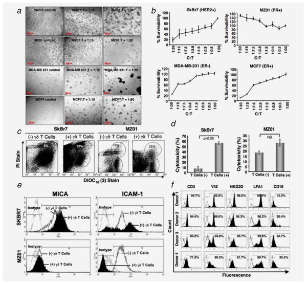
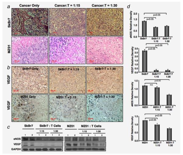
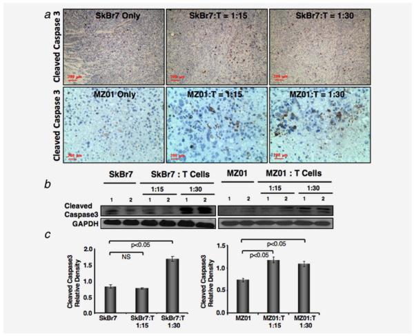
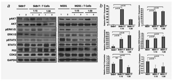
Innate immune system has been known to play an important role in inhibiting the malignant transformation, tumor progression and invasion. However, the mechanistic basis remains ambiguous. Despite polyclonality of human γδ T cells, Vγ2Vδ2 T cell subset was shown to recognize and limit the growth of various tumors at various degrees. The differential recognition of the tumor cells by Vγ2Vδ2 T cells are yet to be defined. Our study reveals that γδ T cells limit in vitro growth of most breast tumor cells, such as SkBr7 (HER2+), MCF7 (ER+) and MDA-MB-231 (ER-) by inhibiting their survival and inducing apoptosis, except BrCa-MZ01 (PR+) cells. To investigate detail mechanisms of antineoplastic effects, we found that cell death was associated with the surface expression levels of MICA/B and ICAM1. Molecular signaling analysis demonstrated that inhibition of cell growth by γδ T cells was associated with the lower expression levels of cell survival-related molecules such as AKT, ERK and concomitant upregulation of apoptosis-related molecules, such as PARP, cleaved caspase 3 and tumor suppressor genes PTEN and P53. However, opposite molecular signaling was observed in the resistant cell line after coculture with γδ T cells. In vivo, antineoplastic effects of γδ T cells were also documented, where tumor growth was inhibited due to the downregulation of survival signals, strong induction of apoptotic molecules, disruption of microvasculature and increased infiltration of tumor associated macrophages. These findings reveal that a complex molecular signaling is involved in γδ T cell-mediated antineoplastic effects.

Keywords: NOD/SCID mice; Vγ2Vδ2 T cell; angiogenesis; apoptosis; breast cancer; cell survival; xenotransplant.


Figures

Figure 1
Figure 1
Morphological changes of breast cancer cells after incubation with γδ T cells. (a) Various human breast cancer cell lines (SkBr7, BrCa-MZ01, MDA-MB-231 and MCF7) were cocultured with ex vivo expanded γδ T cells for 24 hr with a ratio of cancer: γδ T cells, C:T = 1:0; 1:15; 1:30 and the morphology of cells was imaged under a phase contrast microscope. (b) Breast cancer cell lines (SkBr7, BrCa-MZ01, MDA-MB-231 and MCF7) were cocultured with various ratios of γδ T cells for 24 hr. MTT assay was performed to evaluate tumor cell survival and proliferation after gentle removal of γδ T cells. (c) Cytotoxicity of breast cancer cells in presence of γδ T cells. Breast cancer cell lines (SkBr7, BrCa-MZ01) were cocultured with various ratios of γδ T cells for 24 hr. Flowcytometric analysis was performed to evaluate breast cancer cell cytotoxicity induced by γδ T cells after 24 hr of coculture using a LIVE/DEAD cell-mediated viability/cytotoxicity kit. In brief, breast cancer cells were labeled with DiOC18 (3), washed and then exposed to γδ T cells for 24 hr. After 24 hr, cells were labeled with propidium iodide (PI) and then subjected to flowcytometric evaluation. (d) Graphical presentation of cumulative cytotoxic lysis (%) of SkBr7 and BrCa-MZ01 cells of three independent experiments using γδ T cells. (e) The surface expression of molecules on breast cancer cells in presence or absence of γδ T cells was assessed by flowcytometry. SkBr7 and BrCa-MZ01 cells were cocultured with γδ T cells for 24 hr at 1:10 ratios and assessed for MICA and ICAM1 levels, isotype AB was used as a control. (f) Flow cytometric analysis of ex vivo expanded γδ T cells from four different healthy donors at Day 17 of ex vivo expansion. Each experiment was performed at least three times using γδ T cells from 3–4 donors. [Color figure can be viewed in the online issue, which is available at wileyonlinelibrary.com.]
Figure 2
Figure 2
Expression of signaling molecules on breast cancer cells in presence or absence of γδ T cells. Breast cancer cells were cocultured with γδ T cells for 24 hr at 1:5 ratios. Total proteins were isolated from cancer cells after gentle removal of γδ T cells, and Western blot was performed for various signaling molecules stated in SkBr7 (left panel) and BrCa-MZ01 (right panel) cells. Each experiment was performed at least three times using γδ T cells from three different donors.
Figure 3
Figure 3
In vivo antitumorigenic effects of γδ T cells. Human breast cancer cells such as (a) SkBr7 or, (c) BrCa-MZ01 were coinjected subcutaneously with γδ T cells (with a ratio of Cancer:T = 1:15 or 1:30), cancer cells alone or γδ T cells alone (30 million/mouse) in immunocompromised NOD/SCID male mice, and images were presented after 4 weeks of tumor growth. (b and d) After 4 weeks of xenotransplant tumor volume was measured. Reduced tumor size and weight were observed in the mice, which received coinjection of γδ T cells compared to breast cancer cell alone. No tumors were detected in animals xenotransplanted with γδ T cells alone. n = 3–5 mice per group and used γδ T cells from three different donors. [Color figure can be viewed in the online issue, which is available at wileyonlinelibrary.com.]
Figure 4
Figure 4
Immunopathology of xenograft tumors. (a) Lower number of microvessels was observed in SkBr7 or BrCa-MZ01 cell-mediated tumors, which were coinjected with γδ T cells (with a ratio of Cancer:T = 1:15 or 1:30), compared to SkBr7 or BrCa-MZ01 cells alone. The size of the cells was smaller in BrCa-MZ01-mediated tumors, which were coinjected with γδ T cells compared to BrCa-MZ01 cells alone (lower panels). (b) Angiogenesis was evaluated in xenografts of breast cancer cells (SkBr7 or BrCa-MZ01) coinjected with γδ T cells by using anti VEGF Ab. Decreased levels of VEGF expression were observed in both cancer cell xenografts after coinjection of γδ T cells at either concentration, compared to cancer cells alone. (c) Level of angiogenesis-related signaling molecules (VEGF and eNOS) was evaluated using western blot. (d) Western blot protein expression levels of VEGF and eNOS molecules were measured by densitometric scanning using UN-SCAN-IT software (Silk Scientific) and graphically presented. Each experiment was performed three times. NS = nonsignificant. [Color figure can be viewed in the online issue, which is available at wileyonlinelibrary.com.]
Figure 5
Figure 5
Increased apoptosis in tumors coinjected with γδ T cells. (a) Immunohistochemical analysis of xenografts of breast cancer cells (SkBr7 or BrCa-MZ01) coinjected with γδ T cells was performed using antiapoptotic marker (cleaved Caspase 3). Increased levels of cleaved Caspase 3 staining were observed in both xenografts after coinjection of γδ T cells at either concentration, compared to cancer cells alone. (b) Cleaved Caspase 3 level was analyzed by western blot from the xenografts. Cleaved Caspase 3 level was increased in tumors of mice that received breast cancer cells coinjected with γδ T cells, compared to cancer cells alone. (c) Western blot protein expression level of cleaved Caspase 3 molecule was measured by densitometric scanning using UN-SCAN-IT software (Silk Scientific) and graphically presented. Each experiment was performed three times. NS = nonsignificant. [Color figure can be viewed in the online issue, which is available at wileyonlinelibrary.com.]
Figure 6
Figure 6
Decreased levels of survival genes in xenograft tumors coinjected with γδ T cells. (a) Western blot analysis of survival signaling molecules such as AKT, ERK, STAT3 was performed in tumors of SkBr7 (left panel), and BrCa-MZ01 (right panel) of mice that received breast cancer cells along with γδ T cells or cancer cells alone. (b) Effect of γδ T cells on the TAM in tumor xenografts. Significant increase in the expression of macrophage chemotactic protein1 (MCP1) in tumors coinjected with γδ T cells was observed compared to tumors alone. Significant increase in the expression of inducible nitric oxide synthase (iNOS), signature of M2 tumor suppressive macrophages in tumors coinjected with γδ T cells was observed compared to tumors alone. Significant decrease in the expression of Arginase1 (Arg1), signature of M1 tumor-promoting macrophages in tumors coinjected with γδ T cells was observed compared to tumors alone. Each experiment was performed three times. NS = nonsignificant.

References

    1. Jemal A, Bray F, Center MM, et al. Global cancer statistics. CA Cancer J Clin. 2011;61:69–90. - PubMed
    1. Wu Y, Mohamed H, Chillar R, et al. Clinical significance of Akt and HER2/neu overexpression in African-American and Latina women with breast cancer. Breast Cancer Res. 2008;10:R3. - PMC - PubMed
    1. Taneja P, Maglic D, Kai F, et al. Classical and novel prognostic markers for breast cancer and their clinical significance. Clin Med Insights Oncol. 2010;4:15–34. - PMC - PubMed
    1. Morita CT, Parker CM, Brenner MB, et al. TCR usage and functional capabilities of human gamma delta T cells at birth. J Immunol. 1994;153:3979–88. - PubMed
    1. Alam SM, Clark JS, Leech V, et al. T cell receptor gamma/delta expression on lymphocyte populations of breast cancer patients. Immunol Lett. 1992;31:279–83. - PubMed

Publication types

Substances