. 2013:12:241-78.
doi: 10.1007/978-94-007-5561-1_8.

The iron metallome in eukaryotic organisms

Affiliations

The iron metallome in eukaryotic organisms

Adrienne C Dlouhy et al. Met Ions Life Sci. 2013.

Abstract

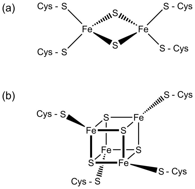
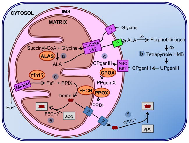
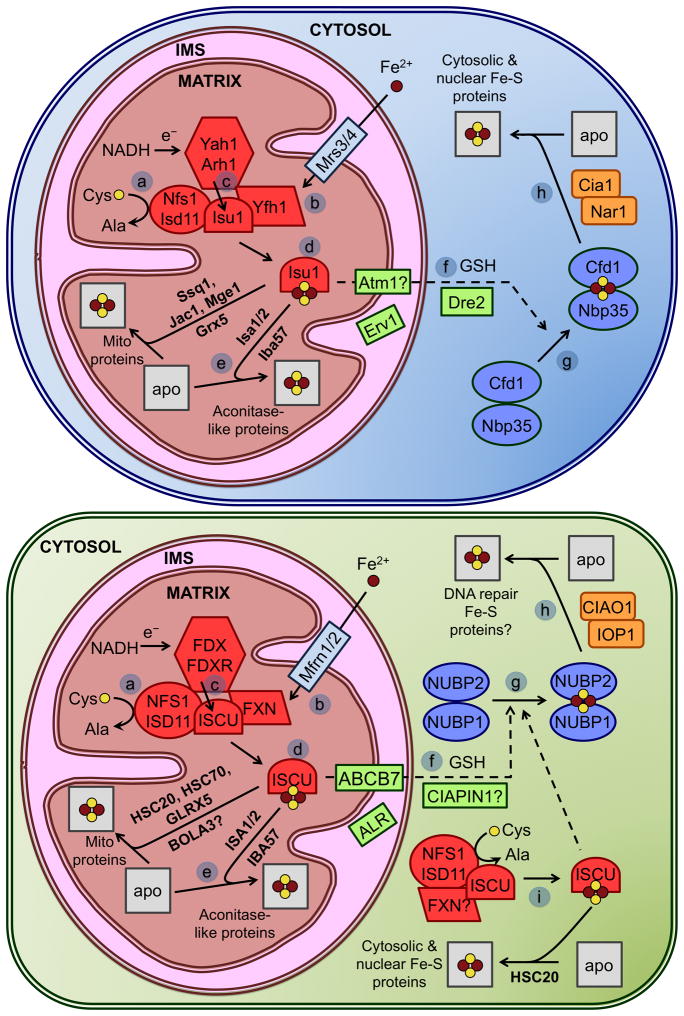
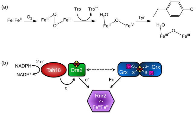
This chapter is focused on the iron metallome in eukaryotes at the cellular and subcellular level, including properties, utilization in metalloproteins, trafficking, storage, and regulation of these processes. Studies in the model eukaryote Saccharomyces cerevisiae and mammalian cells will be highlighted. The discussion of iron properties will center on the speciation and localization of intracellular iron as well as the cellular and molecular mechanisms for coping with both low iron bioavailability and iron toxicity. The section on iron metalloproteins will emphasize heme, iron-sulfur cluster, and non-heme iron centers, particularly their cellular roles and mechanisms of assembly. The section on iron uptake, trafficking, and storage will compare methods used by yeast and mammalian cells to import iron, how this iron is brought into various organelles, and types of iron storage proteins. Regulation of these processes will be compared between yeast and mammalian cells at the transcriptional, post-transcriptional, and post-translational levels.


Figures

Figure 1
Figure 1
Mössbauer spectra comparing differing iron species found in (a) respiring mitochondria, (b) fermenting mitochondria, (c) Atm1-depleted mitochondria, and (d) whole fermenting yeast cells. (reprinted with permission from ref [8])
Figure 2
Figure 2
Structural comparison of Fe3+-binding siderophore moieties: (a) catechol and (b) 2,5-DHBA found in mammalian cells, and (c) 2,3-DHBA found in bacterial enterobactin.
Figure 3
Figure 3
Proposed structure of the glutathione-coordinating complex with Fe(II) found in the labile iron pool.
Figure 4
Figure 4
Heme cofactor. Structure of protoporphyrin IX with ferrous iron inserted.
Figure 5
Figure 5
Common forms of iron-sulfur cluster cofactors: (a) [2Fe-2S] and (b) [4Fe-4S].
Figure 6
Figure 6
Heme biosynthesis pathway in eukaryotes. (a) Glycine is transported into the mitochondrial matrix via an unknown mechanism where it is combined with succinyl-CoA by ALA synthase (ALAS) to form ALA. (b) ALA is transported out to the cytosol where it is converted to CPgenIII through four conserved steps. (c) ABCB6 is the transporter proposed to import CPgenIII to the IMS where it is converted first to PPgenIX by CPOX, then to protoporphyrin IX (PPIX) by PPOX. (d) PPIX is transported to the matrix where iron is inserted by ferrochelatase (FECH). The proposed Fe(II) importers are Mrs3/4 (Mfrn1/2 in mammalian cells). (e) Assembled heme is inserted into target apo proteins to form hemoproteins in the mitochondria, possibly aided by FECH. (f) Heme is inserted into target apo proteins in the cytosol, possibly aided by GSTs.
Figure 7
Figure 7
Mitochondrial and cytosolic Fe-S cluster assembly in yeast (top panel) and mammalian cells (bottom panel). (a) In the mitochondria, sulfur is obtained from the cysteine desulfurase Nfs1, interacting with Isd11. Nfs1 transfers sulfur as a persulfide to Isu1/2 (ISCU in humans). (b) Iron is imported to the mitochondria by the transporters Mrs3/4 (mitoferrins or Mfrn1/2 in humans) and possibly donated to Isu1/2 through frataxin (FXN, or Yfh1 in yeast). (c) Electrons are donated by NADH through the ferredoxin-ferredoxin reductase pair Yah1-Arh1 to reduce S0 to S2−. (d) The assembled Fe-S cluster is transferred to target proteins by a chaperone system consisting of Ssq1, Jac1, Mge1, and Grx5 (HSC20, HSC70, and GLRX5 in humans). (e) Isa1/2 specifically delivers clusters to aconitase-like proteins. (f) An unknown substrate produced by the ISC machinery is exported out to the cytosol by the transporter Atm1 (ABCB7 in humans). This process may also include the sulfhydryl oxidase Erv1 (ALR in humans), GSH, and Dre2 (CIAPIN1). (g) Fe-S clusters are assembled in the cytosol on the scaffold complex formed by Cfd1 and Nbp35 (NUBP2 and NUBP1 in humans, respectively). (h) The assembled cluster is transferred to target cytosolic and nuclear proteins by Nar1 and Cia1 (IOP1 and CIAO1 in humans). (i) Mammalian cells also express cytosolic versions of NFS1, ISD11, ISCU, HSC20, and possibly FXN that may also facilitate de novo assembly of Fe-S clusters outside the mitochondria.
Figure 8
Figure 8
Formation of the class Ia RNR diferric-tyrosyl radical cofactor. (a) Formation reaction for the diferric-tyrosyl radical cofactor. (b) Model for the biosynthesis pathway of active Rnr2. Tah18 and Dre2 are proposed to donate electrons for diferric-tyrosyl radical formation on Rnr2. The [2Fe-2S] Grx3/4 homodimer most likely donates iron for the cofactor. Reducing equivalents from Dre2 may also be required for iron delivery (dashed line).
Figure 9
Figure 9
Iron uptake systems in S. cerevisiae. Arn1, Sit1, Enb1, and Taf1 are transporters for Fe3+-siderophore complexes. The ferrireductases Fre1/2 reduce environmental Fe3+ to Fe2+. Fet3 and Ftr1 form the high-affinity iron uptake system. Fet3 oxidizes Fe2+ to Fe3+ and Ftr1 transports this across the plasma membrane. Fet4 is a low-affinity transporter responsible for uptake under iron-replete conditions. Smf1, a member of the NRAMP family of transporters, is a H+/M+ symporter for some transition metals like Fe2+, Mn2+, and Zn2+. Aft1 and Aft2 are transcriptional regulators that activate expression of the iron regulon, including these iron uptake systems, under low iron conditions.
Figure 10
Figure 10
Iron import and export in a generic mammalian cell. The plasma protein transferrin (Tf) binds 2 ferric ions for uptake by the transferrin receptors (TfR1 and TfR2). TfR1 is found in all cell types, while TfR2 is limited to liver, intestinal, and red blood cells. Tf(Fe3+)2-bound TfR is internalized by endocytosis and ferric iron is released in the acidic environment of the endosome. Tf and TfR are recycled to the plasma membrane. DMT1 is involved in release of Tf iron from the endosome following reduction of Fe3+ to Fe2+ by STEAP ferrireductases. Dcytb (cytochrome b-like ferrireductase) reduces dietary Fe3+ to Fe2+, which is imported by DMT1 at the plasma membrane. The plasma proteins haptoglobin and hemopexin bind hemoglobin and free heme, respectively, produced by erythrocyte destruction. Haptoglobin-hemoglobin and heme-hemopexin complexes are recognized by CD163 and CD91 receptors, respectively, for subsequent endocytosis. Heme is also imported via HCP1 and HRG1. HO-1 (heme oxygenase-1) catalyzes degradation of the heme to remove iron. Imported iron can be stored in ferritin or trafficked to the mitochondria for synthesis of heme and Fe-S clusters. FPN is the iron exporter, transporting Fe2+ out of the cell. Ceruloplasmin (Cp) oxidizes this Fe2+ to Fe3+ for binding to Tf.
Figure 11
Figure 11
mRNA regulation of iron trafficking and utilization factors by the IRE-IRP system. In iron-deplete cells, IRP1 lacks the [4Fe-4S] and IRP2 is stabilized, thus both IRPs are able to bind the target IREs. Translation of mRNAs with 5′ IREs (e.g. ferritin) is blocked, while mRNAs with 3′ IREs (e.g. TfR1) are stabilized and translated. In iron-replete cells, IRP1 binds a [4Fe-4S] cluster that precludes IRE binding, while IRP2 is degraded by the proteasome. IREs of the target RNAs are unoccupied. mRNAs with IREs in the 5′ UTR are translated, while RNase degrades mRNAs with IREs in the 3′ UTR.
Figure 12
Figure 12
Proposed model for regulation of Aft1/2 in S. cerevisiae. Under iron replete conditions (left panel), the [2Fe-2S]-bridged Fra2-Grx3/4 complex (and possibly Fra1) relays the cellular iron status to Aft1/2, leading to Aft1/2 oligomerization. Aft1/2 is consequently shuttled to the cytosol by the exportin Msn5, deactivating the iron regulon. Under low iron conditions (right panel), Grx3/4 and Fra2 cannot bind an Fe-S cluster, and Aft1/2 does not receive the Fe-S signal. Msn5 does not recognize Aft1/2 and it accumulates in the nucleus where it activates the iron regulon.

References

    1. Eide DJ, Clark S, Nair TM, Gehl M, Gribskov M, Guerinot ML, Harper JF. Genome Biol. 2005;6:R77. - PMC - PubMed
    1. Epsztejn S, Glickstein H, Picard V, Slotki IN, Breuer W, Beaumont C, Cabantchik ZI. Blood. 1999;94:3593–3603. - PubMed
    1. Petrat F, Rauen U, de Groot H. Hepatology. 1999;29:1171–1179. - PubMed
    1. Ceccarelli D, Gallesi D, Giovannini F, Ferrali M, Masini A. Biochem Biophys Res Commun. 1995;209:53–59. - PubMed
    1. Gao X, Qian M, Campian JL, Marshall J, Zhou Z, Roberts AM, Kang YJ, Prabhu SD, Sun XF, Eaton JW. Free Radic Biol Med. 2010;49:401–407. - PMC - PubMed