. 2013 Apr 17;33(16):7020-6.
doi: 10.1523/JNEUROSCI.2325-12.2013.

Sodium channel cleavage is associated with aberrant neuronal activity and cognitive deficits in a mouse model of Alzheimer's disease

Affiliations

Sodium channel cleavage is associated with aberrant neuronal activity and cognitive deficits in a mouse model of Alzheimer's disease

Brian F Corbett et al. J Neurosci. .

Erratum in

  • J Neurosci. 2013 Jun 26;33(26):10934. Brown, Jon T [added]; Randall, Andrew D [added]

Abstract

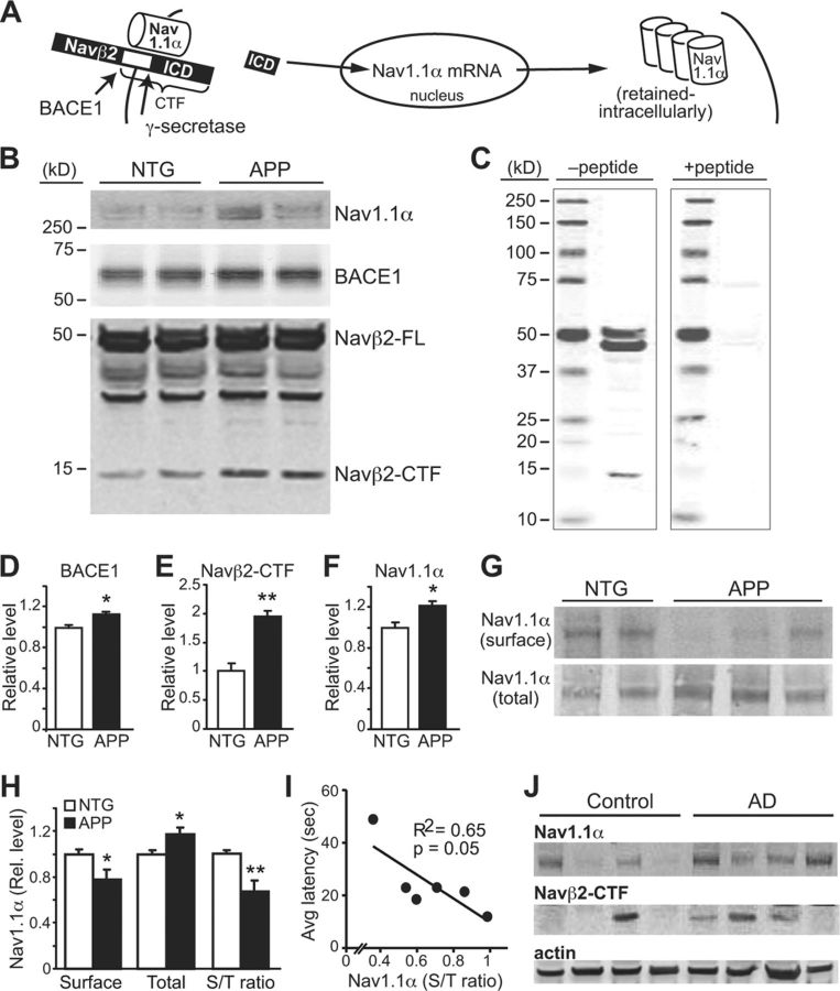
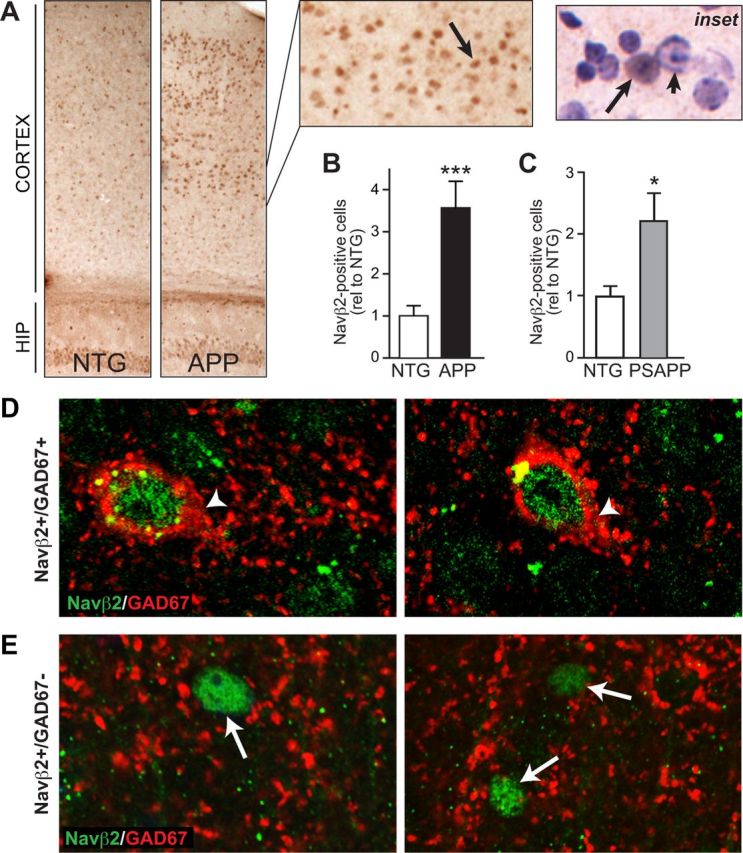
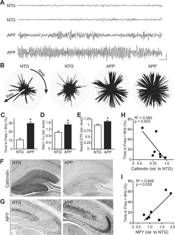
BACE1 is the rate-limiting enzyme that cleaves amyloid precursor protein (APP) to produce the amyloid β peptides that accumulate in Alzheimer's disease (AD). BACE1, which is elevated in AD patients and APP transgenic mice, also cleaves the β2-subunit of voltage-gated sodium channels (Navβ2). Although increased BACE1 levels are associated with Navβ2 cleavage in AD patients, whether Navβ2 cleavage occurs in APP mice had not yet been examined. Such a finding would be of interest because of its potential impact on neuronal activity: previous studies demonstrated that BACE1-overexpressing mice exhibit excessive cleavage of Navβ2 and reduced sodium current density, but the phenotype associated with loss of function mutations in either Navβ-subunits or pore-forming α-subunits is epilepsy. Because mounting evidence suggests that epileptiform activity may play an important role in the development of AD-related cognitive deficits, we examined whether enhanced cleavage of Navβ2 occurs in APP transgenic mice, and whether it is associated with aberrant neuronal activity and cognitive deficits. We found increased levels of BACE1 expression and Navβ2 cleavage fragments in cortical lysates from APP transgenic mice, as well as associated alterations in Nav1.1α expression and localization. Both pyramidal neurons and inhibitory interneurons exhibited evidence of increased Navβ2 cleavage. Moreover, the magnitude of alterations in sodium channel subunits was associated with aberrant EEG activity and impairments in the Morris water maze. Together, these results suggest that altered processing of voltage-gated sodium channels may contribute to aberrant neuronal activity and cognitive deficits in AD.


Figures

Figure 1.
Figure 1.
Voltage-gated sodium channel alterations in APP mice and AD patients. A, Cleavage of Navβ2 by BACE1 and γ-secretase and alterations in Nav1.1α. Modified from Kim et al. (2007). B, Increased BACE1 expression and Navβ2-CTFs in cortical lysates from 5- to 6-month-old APP compared with NTG mice (middle and bottom). Navβ2-CTFs are further cleaved by γ-secretase to produce an ICD that translocates to the nucleus to increase Nav1.1α expression, which is also increased in APP mice (top). C, Incubation of the anti-Navβ2 antibody with blocking peptide eliminated the bands around 50 and 13 kDa, indicating that these bands are Navβ2-specific bands. D–F, Quantification of blots in B, n = 8–10/genotype. G, Reduced surface levels of Nav1.1α in APP mice. Cortical slices were biotinylated and immunoprecipated for cell-surface proteins. APP mice had less surface Nav1.1α despite increased total levels, quantified in H, n = 6/genotype. I, Relationship between surface levels of Nav1.1α and performance in the hidden platform portion of the water maze, a hippocampal-dependent memory task. J, Cortical lysates from AD patients also exhibit increased Navβ2-CTFs and total Nav1.1α levels, similar to previously published (Kim et al., 2007). *p < 0.05, **p < 0.01.
Figure 2.
Figure 2.
Increased numbers of Navβ2-positive nuclei in cortex of APP mice. Navβ2-positive nuclei were assessed in brain sections from APP mice after immunohistochemical staining with an antibody that recognizes Navβ2-ICD. A, APP mice exhibit increased numbers of Navβ2-positive nuclei in cortical regions. Arrow points to a Navβ2-positive nucleus. Inset, Hematoxylin counterstain demonstrates nuclear Navβ2 localization (arrow). Arrowhead, Nucleus with no Navβ2. B, Quantification of nuclear Navβ2 staining in APP and NTG mice, n = 7–9/genotype. C, Quantification of Navβ2 staining in brain sections from PSAPP mice reveals a significant increase in Navβ2-positive nuclei also in this mouse model, n = 5–7/genotype. D, E, Double-labeling of Navβ2 and GAD67 in brain sections from APP mice. Confocal images of Navβ2 and GAD67 immunostaining demonstrate that some Navβ2-positive nuclei are found in cells that also express GAD67 (D, arrowheads), but others do not express GAD67 (E, arrows). *p < 0.05, ***p < 0.001.
Figure 3.
Figure 3.
Aberrant neuronal activity in APP mice. A, EEG traces demonstrate increased amplitude and frequency of brain activity in two representative NTG and two APP mice at 5–7 months of age. B, Radar plots show time (2 h) as a continuous parameter, and frequency from 0 to 10 Hz in concentric circles. C–E, APP mice exhibit longer durations of higher frequency activity than NTG mice (C), which is associated with increased total Nav1.1α levels (D) and increased Navβ2 cleavage (E) (n = 7–9/genotype). F, G, APP mice exhibit decreased calbindin expression in dentate gyrus (F) and ectopic NPY expression in mossy fibers (G). H, I, Calbindin expression in the dentate gyrus correlated inversely (H) and NPY expression in mossy fibers correlated positively (I) with the EEG parameter of time spent in high-frequency. *p < 0.05.

References

    1. Amatniek JC, Hauser WA, DelCastillo-Castaneda C, Jacobs DM, Marder K, Bell K, Albert M, Brandt J, Stern Y. Incidence and predictors of seizures in patients with Alzheimer's disease. Epilepsia. 2006;47:867–872. doi: 10.1111/j.1528-1167.2006.00554.x. - DOI - PubMed
    1. Bakker A, Krauss GL, Albert MS, Speck CL, Jones LR, Stark CE, Yassa MA, Bassett SS, Shelton AL, Gallagher M. Reduction of hippocampal hyperactivity improves cognition in amnestic mild cognitive impairment. Neuron. 2012;74:467–474. doi: 10.1016/j.neuron.2012.03.023. - DOI - PMC - PubMed
    1. Bertram L, Lill CM, Tanzi RE. The genetics of Alzheimer disease: back to the future. Neuron. 2010;68:270–281. doi: 10.1016/j.neuron.2010.10.013. - DOI - PubMed
    1. Blennow K, de Leon MJ, Zetterberg H. Alzheimer's disease. Lancet. 2006;368:387–403. doi: 10.1016/S0140-6736(06)69113-7. - DOI - PubMed
    1. Bourne KZ, Ferrari DC, Lange-Dohna C, Rossner S, Wood TG, Perez-Polo JR. Differential regulation of BACE1 promoter activity by nuclear factor-kappaB in neurons and glia upon exposure to beta-amyloid peptides. J Neurosci Res. 2007;85:1194–1204. doi: 10.1002/jnr.21252. - DOI - PubMed

Publication types

MeSH terms

LinkOut - more resources