. 2013 Apr 17;33(16):7050-6.
doi: 10.1523/JNEUROSCI.3081-12.2013.

Application of high-frequency repetitive transcranial magnetic stimulation to the DLPFC alters human prefrontal-hippocampal functional interaction

Affiliations

Application of high-frequency repetitive transcranial magnetic stimulation to the DLPFC alters human prefrontal-hippocampal functional interaction

Edda Bilek et al. J Neurosci. .

Abstract

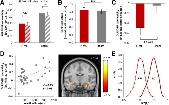
Neural plasticity is crucial for understanding the experience-dependent reorganization of brain regulatory circuits and the pathophysiology of schizophrenia. An important circuit-level feature derived from functional magnetic resonance imaging (fMRI) is prefrontal-hippocampal seeded connectivity during working memory, the best established intermediate connectivity phenotype of schizophrenia risk to date. The phenotype is a promising marker for the effects of plasticity-enhancing interventions, such as high-frequency repetitive transcranial magnetic stimulation (rTMS), and can be studied in healthy volunteers in the absence of illness-related confounds, but the relationship to brain plasticity is unexplored. We recruited 39 healthy volunteers to investigate the effects of 5 Hz rTMS on prefrontal-hippocampal coupling during working memory and rest. In a randomized and sham-controlled experiment, neuronavigation-guided rTMS was applied to the right dorsolateral prefrontal cortex (DLPFC), and fMRI and functional connectivity analyses [seeded connectivity and psychophysiological interaction (PPI)] were used as readouts. Moreover, the test-retest reliability of working-memory related connectivity markers was evaluated. rTMS provoked a significant decrease in seeded functional connectivity of the right DLPFC and left hippocampus during working memory that proved to be relatively time-invariant and robust. PPI analyses provided evidence for a nominal effect of rTMS and poor test-retest reliability. No effects on n-back-related activation and DLPFC-hippocampus resting-state connectivity were observed. These data provide the first in vivo evidence for the effects of plasticity induction on human prefrontal-hippocampal network dynamics, offer insights into the biological mechanisms of a well established intermediate phenotype linked to schizophrenia, and underscores the importance of the choice of outcome measures in test-retest designs.


Figures

Figure 1.
Figure 1.
A, Spatial consistency of rTMS target sites in the right DLPFC. For illustration purposes, individual coordinates of the center of stimulation sites in DLPFC were derived from the functional neuronavigation software, approximated by 5 mm spheres, normalized to standard MNI space, and superimposed to create a heat map of TMS stimulation sites for all subjects. The group map is overlaid on sagittal (left) and transversal (right) sections of a structural template. Color bar represents number of subjects with overlapping stimulation targets at a given voxel in normalized space. Please note that the figure underestimates the topology of locally induced electrical fields. B, Effects of rTMS on DLPFC–hippocampus functional coupling as quantified with a correlative measure (“seeded connectivity”). Left, Significant decrease in functional connectivity of the right DLPFC and left hippocampus under rTMS relative to sham TMS during working memory performance, but no significant effects at rest. Bar plots depict the mean value for the connectivity estimates of the peak voxel stratified by treatment condition. Error bars illustrate the variance of parameter estimates (±SEM). Right, heat map of the n-back group connectivity statistics overlaid on a coronal template section of the brain (p = 0.005 uncorrected, for presentation purposes). Color bar represents t values. SC, Seeded connectivity; HF, hippocampus formation; NS, not significant.
Figure 2.
Figure 2.
A, No significant main effect of time or rTMS condition by time interaction effect on DLPFC–hippocampus seeded connectivity in a split-half analysis of the n-back data (p > 0.05, see results section for details). B, No significant effects of rTMS on working-memory related brain activations in DLPFC in the area under the coil (p > 0.05, see Results for details). The bar plot depicts parameter estimates derived from individual stimulation sites in native space. C, Nominal significant effect of rTMS on DLPFC–hippocampus functional connectivity as quantified with PPI. Bar plot depicts the mean contrast estimates derived from nominally significant clusters (p < 0.05, uncorrected, see Results for details). All error bars illustrate the variance of parameter estimates (±SEM). D, Significant association of DLPFC–hippocampus seeded connectivity estimates with n-back-related reaction times in the sham condition (p = 0.05, corrected, see Results for details). The heat map depicts the corresponding group statistic on a coronal template section of the brain (p = 0.005 uncorrected, for presentation purposes). Color bar represents t values. E, Test-retest reliability of n-back-related DLPFC–hippocampus connectivity estimates. The graph depicts ICC (2,1) density plots across hippocampus voxels for PPI (dashed line) and seeded connectivity (solid line) estimates.

References

    1. Bennett CM, Miller MB. How reliable are the results from functional magnetic resonance imaging? Ann N Y Acad Sci. 2010;1191:133–155. doi: 10.1111/j.1749-6632.2010.05446.x. - DOI - PubMed
    1. Bestmann S, Baudewig J, Siebner HR, Rothwell JC, Frahm J. Subthreshold high-frequency TMS of human primary motor cortex modulates interconnected frontal motor areas as detected by interleaved fMRI-TMS. Neuroimage. 2003;20:1685–1696. doi: 10.1016/j.neuroimage.2003.07.028. - DOI - PubMed
    1. Bosch M, Hayashi Y. Structural plasticity of dendritic spines. Curr Opin Neurobiol. 2012;22:383–388. doi: 10.1016/j.conb.2011.09.002. - DOI - PMC - PubMed
    1. Callicott JH, Egan MF, Mattay VS, Bertolino A, Bone AD, Verchinksi B, Weinberger DR. Abnormal fMRI response of the dorsolateral prefrontal cortex in cognitively intact siblings of patients with schizophrenia. Am J Psychiatry. 2003;160:709–719. doi: 10.1176/appi.ajp.160.4.709. - DOI - PubMed
    1. Cashdollar N, Malecki U, Rugg-Gunn FJ, Duncan JS, Lavie N, Duzel E. Hippocampus-dependent and -independent theta-networks of active maintenance. Proc Natl Acad Sci U S A. 2009;106:20493–20498. doi: 10.1073/pnas.0904823106. - DOI - PMC - PubMed

Publication types

MeSH terms

LinkOut - more resources