

Altered splicing of ATP6AP2 causes X-linked parkinsonism with spasticity (XPDS)
- PMID: 23595882
- PMCID: PMC3723311
- DOI: 10.1093/hmg/ddt180
Altered splicing of ATP6AP2 causes X-linked parkinsonism with spasticity (XPDS)
Abstract
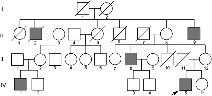
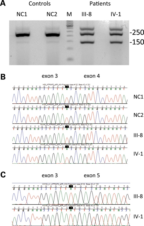
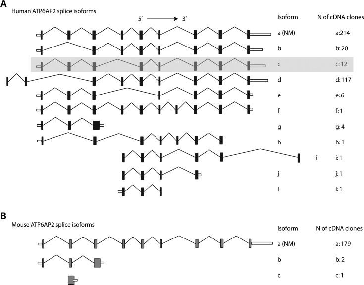
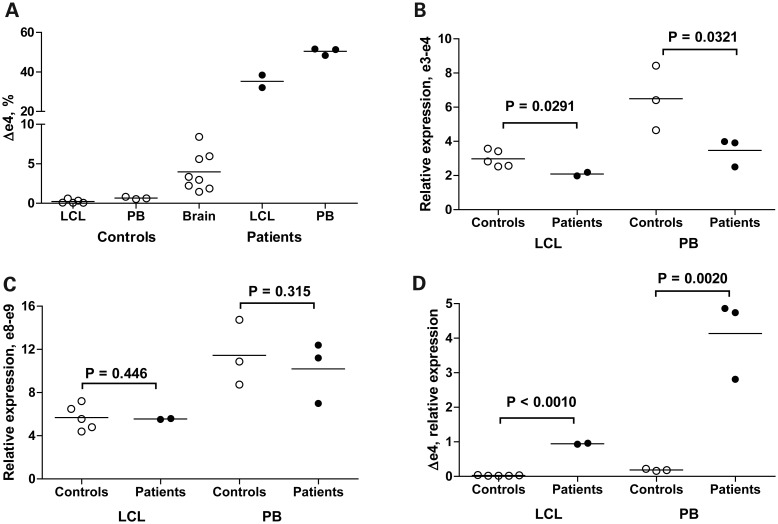
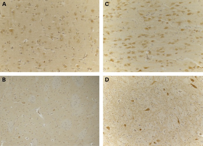
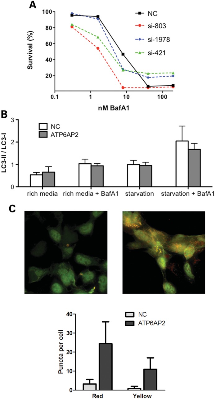
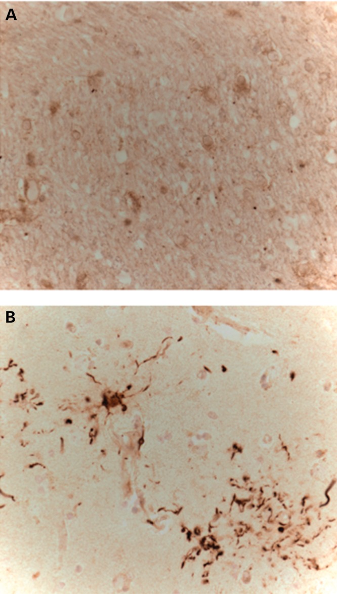
We report a novel gene for a parkinsonian disorder. X-linked parkinsonism with spasticity (XPDS) presents either as typical adult onset Parkinson's disease or earlier onset spasticity followed by parkinsonism. We previously mapped the XPDS gene to a 28 Mb region on Xp11.2-X13.3. Exome sequencing of one affected individual identified five rare variants in this region, of which none was missense, nonsense or frame shift. Using patient-derived cells, we tested the effect of these variants on expression/splicing of the relevant genes. A synonymous variant in ATP6AP2, c.345C>T (p.S115S), markedly increased exon 4 skipping, resulting in the overexpression of a minor splice isoform that produces a protein with internal deletion of 32 amino acids in up to 50% of the total pool, with concomitant reduction of isoforms containing exon 4. ATP6AP2 is an essential accessory component of the vacuolar ATPase required for lysosomal degradative functions and autophagy, a pathway frequently affected in Parkinson's disease. Reduction of the full-size ATP6AP2 transcript in XPDS cells and decreased level of ATP6AP2 protein in XPDS brain may compromise V-ATPase function, as seen with siRNA knockdown in HEK293 cells, and may ultimately be responsible for the pathology. Another synonymous mutation in the same exon, c.321C>T (p.D107D), has a similar molecular defect of exon inclusion and causes X-linked mental retardation Hedera type (MRXSH). Mutations in XPDS and MRXSH alter binding sites for different splicing factors, which may explain the marked differences in age of onset and manifestations.
Figures
References
-
- Poorkaj P., Raskind W.H., Leverenz J.B., Matsushita M., Zabetian C.P., Samii A., Kim S., Gazi N., Nutt J.G., Wolff J., et al. A novel X-linked four-repeat tauopathy with Parkinsonism and spasticity. Mov. Disord. 2010;25:1409–1417. doi:10.1002/mds.23085. - DOI - PMC - PubMed
-
- Ng S.B., Bigham A.W., Buckingham K.J., Hannibal M.C., McMillin M.J., Gildersleeve H.I., Beck A.E., Tabor H.K., Cooper G.M., Mefford H.C., et al. Exome sequencing identifies MLL2 mutations as a cause of Kabuki syndrome. Nat. Genet. 2010;42:790–793. doi:10.1038/ng.646. - DOI - PMC - PubMed
-
- Chen Y.Z., Matsushita M.M., Robertson P., Rieder M., Girirajan S., Antonacci F., Lipe H., Eichler E.E., Nickerson D.A., Bird T.D., et al. Autosomal dominant familial dyskinesia and facial myokymia: single exome sequencing identifies a mutation in adenylyl cyclase 5. Arch. Neurol. 2012;69:630–635. doi:10.1001/archneurol.2012.54. - DOI - PMC - PubMed
-
- Cummings C.J., Zoghbi H.Y. Trinucleotide repeats: mechanisms and pathophysiology. Annu. Rev. Genomics Hum. Genet. 2000;1:281–328. doi:10.1146/annurev.genom.1.1.281. - DOI - PubMed
-
- Ludwig J., Kerscher S., Brandt U., Pfeiffer K., Getlawi F., Apps D.K., Schagger H. Identification and characterization of a novel 9.2-kDa membrane sector-associated protein of vacuolar proton-ATPase from chromaffin granules. J. Biol. Chem. 1998;273:10939–10947. doi:10.1074/jbc.273.18.10939. - DOI - PubMed
Publication types
MeSH terms
Substances
Grants and funding
- HL-103010/HL/NHLBI NIH HHS/United States
- HL 1029230/HL/NHLBI NIH HHS/United States
- R01 NS069719/NS/NINDS NIH HHS/United States
- I01 BX000531/BX/BLRD VA/United States
- T32 GM00727/GM/NIGMS NIH HHS/United States
- R01 NS065070/NS/NINDS NIH HHS/United States
- HL 102926/HL/NHLBI NIH HHS/United States
- HL 102924/HL/NHLBI NIH HHS/United States
- P50 AG005136/AG/NIA NIH HHS/United States
- R01NS069719/NS/NINDS NIH HHS/United States
- P50 NS062684/NS/NINDS NIH HHS/United States
- HL-102925/HL/NHLBI NIH HHS/United States
- RC2HG005608/HG/NHGRI NIH HHS/United States
- R01 NS064131/NS/NINDS NIH HHS/United States
- R01NS064131/NS/NINDS NIH HHS/United States
- P01 GM081619/GM/NIGMS NIH HHS/United States
LinkOut - more resources
Full Text Sources
Other Literature Sources
Molecular Biology Databases
Research Materials
Miscellaneous
