t-SNARE Syntaxin2 (STX2) is implicated in intracellular transport of sulfoglycolipids during meiotic prophase in mouse spermatogenesis
- PMID: 23595907
- PMCID: PMC6322434
- DOI: 10.1095/biolreprod.112.107110
t-SNARE Syntaxin2 (STX2) is implicated in intracellular transport of sulfoglycolipids during meiotic prophase in mouse spermatogenesis
Abstract
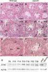
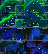
Syntaxin2 (STX2), also known as epimorphin, is a member of the SNARE family of proteins, with expression in various types of cells. We previously identified an ENU-induced mutation, repro34, in the mouse Stx2 gene. The Stx2(repro34) mutation causes male-restricted infertility due to syncytial multinucleation of spermatogenic cells during meiotic prophase. A similar phenotype is also observed in mice with targeted inactivation of Stx2, as well as in mice lacking enzymes involved in sulfoglycolipid synthesis. Herein we analyzed expression and subcellular localization of STX2 and sulfoglycolipids in spermatogenesis. The STX2 protein localizes to the cytoplasm of germ cells at the late pachytene stage. It is found in a distinct subcellular pattern, presumably in the Golgi apparatus of pachytene/diplotene spermatocytes. Sulfoglycolipids are produced in the Golgi apparatus and transported to the plasma membrane. In Stx2(repro34) mutants, sulfoglycolipids are aberrantly localized in both pachytene/diplotene spermatocytes and in multinucleated germ cells. These results suggest that STX2 plays roles in transport and/or subcellular distribution of sulfoglycolipids. STX2 function in the Golgi apparatus and sulfoglycolipids may be essential for maintenance of the constriction between neighboring developing spermatocytes, which ensures ultimate individualization of germ cells in later stages of spermatogenesis.
Keywords: Stx2; glycolipids; meiosis; mouse; spermatogenesis.
Figures


Similar articles
-
A new ENU-induced mutant mouse with defective spermatogenesis caused by a nonsense mutation of the syntaxin 2/epimorphin (Stx2/Epim) gene.J Reprod Dev. 2008 Apr;54(2):122-8. doi: 10.1262/jrd.19186. Epub 2008 Feb 14. J Reprod Dev. 2008. PMID: 18277055
-
HSP70-2 is required for desynapsis of synaptonemal complexes during meiotic prophase in juvenile and adult mouse spermatocytes.Development. 1997 Nov;124(22):4595-603. doi: 10.1242/dev.124.22.4595. Development. 1997. PMID: 9409676
-
The Golgi apparatus of rat pachytene spermatocytes during spermatogenesis.Anat Rec. 1991 Jan;229(1):16-26. doi: 10.1002/ar.1092290104. Anat Rec. 1991. PMID: 1996781
-
Protein expression and cell organelle behavior in spermatogenic cells.Kaibogaku Zasshi. 2001 Jun;76(3):267-79. Kaibogaku Zasshi. 2001. PMID: 11494512 Review.
-
Biosynthesis and biological function of sulfoglycolipids.Proc Jpn Acad Ser B Phys Biol Sci. 2013;89(4):129-38. doi: 10.2183/pjab.89.129. Proc Jpn Acad Ser B Phys Biol Sci. 2013. PMID: 23574804 Free PMC article. Review.
Cited by
-
Genetic defects in human azoospermia.Basic Clin Androl. 2019 Apr 23;29:4. doi: 10.1186/s12610-019-0086-6. eCollection 2019. Basic Clin Androl. 2019. PMID: 31024732 Free PMC article.
-
Genetics of Azoospermia.Int J Mol Sci. 2021 Mar 23;22(6):3264. doi: 10.3390/ijms22063264. Int J Mol Sci. 2021. PMID: 33806855 Free PMC article. Review.
-
Properties, metabolism and roles of sulfogalactosylglycerolipid in male reproduction.Prog Lipid Res. 2018 Oct;72:18-41. doi: 10.1016/j.plipres.2018.08.002. Epub 2018 Aug 25. Prog Lipid Res. 2018. PMID: 30149090 Free PMC article. Review.
-
Expression, sorting, and segregation of Golgi proteins during germ cell differentiation in the testis.Mol Biol Cell. 2015 Nov 5;26(22):4015-32. doi: 10.1091/mbc.E14-12-1632. Epub 2015 Mar 25. Mol Biol Cell. 2015. PMID: 25808494 Free PMC article.
-
Birth of mice from meiotically arrested spermatocytes following biparental meiosis in halved oocytes.EMBO Rep. 2022 Jul 5;23(7):e54992. doi: 10.15252/embr.202254992. Epub 2022 May 19. EMBO Rep. 2022. PMID: 35587095 Free PMC article.
References
-
- Handel MA, Lessard C, Reinholdt L, Schimenti J, Eppig JJ.. Mutagenesis as an unbiased approach to identify novel contraceptive targets. Mol Cell Endocrinol 2006; 250:201–205. - PubMed
-
- Akiyama K, Akimaru S, Asano Y, Khalaj M, Kiyosu C, Masoudi AA, Takahashi S, Katayama K, Tsuji T, Noguchi J, Kunieda T.. A new ENU-induced mutant mouse with defective spermatogenesis caused by a nonsense mutation of the syntaxin 2/epimorphin (Stx2/Epim) gene. J Reprod Dev 2008; 54:122–128. - PubMed
-
- Albertson R, Riggs B, Sullivan W.. Membrane traffic: a driving force in cytokinesis. Trends Cell Biol 2005; 15:92–101. - PubMed
Publication types
MeSH terms
Substances
Grants and funding
LinkOut - more resources
Full Text Sources
Other Literature Sources
Molecular Biology Databases

