

Ligand-bound thyroid hormone receptor contributes to reprogramming of pancreatic acinar cells into insulin-producing cells
- PMID: 23595988
- PMCID: PMC3668771
- DOI: 10.1074/jbc.M112.438192
Ligand-bound thyroid hormone receptor contributes to reprogramming of pancreatic acinar cells into insulin-producing cells
Abstract
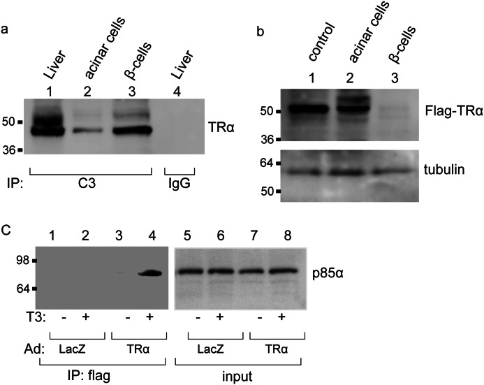
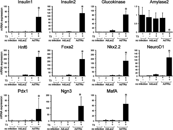
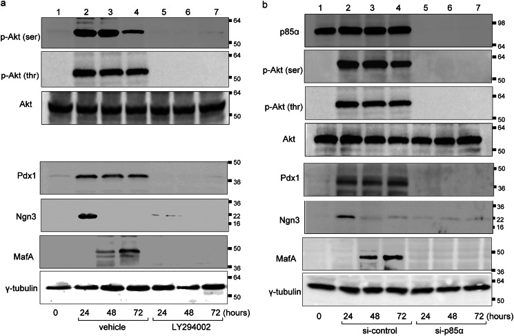
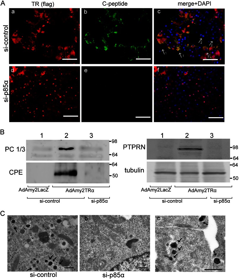
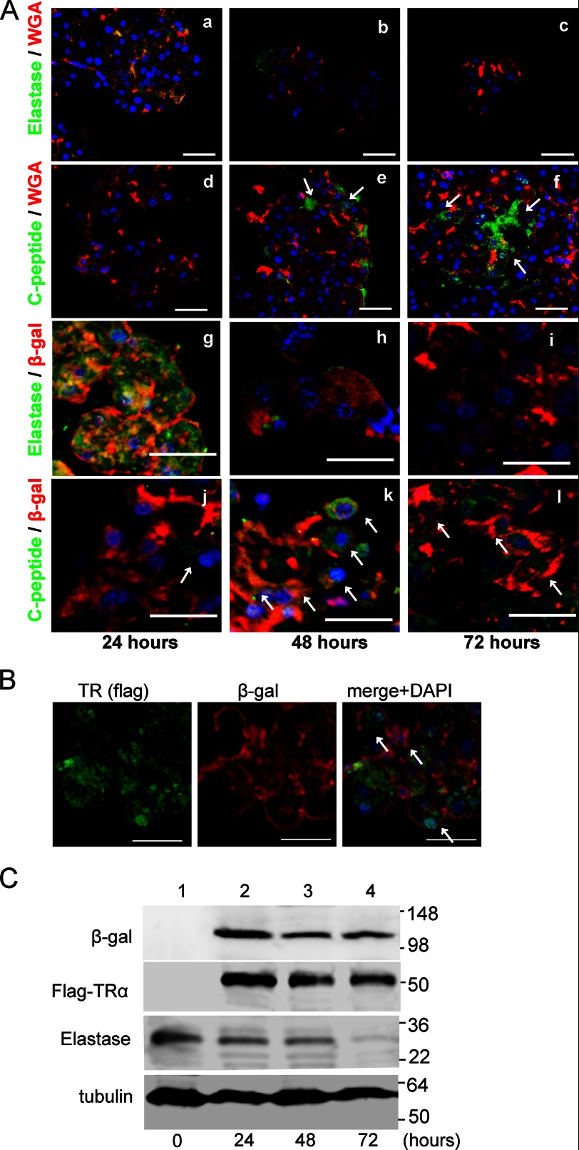
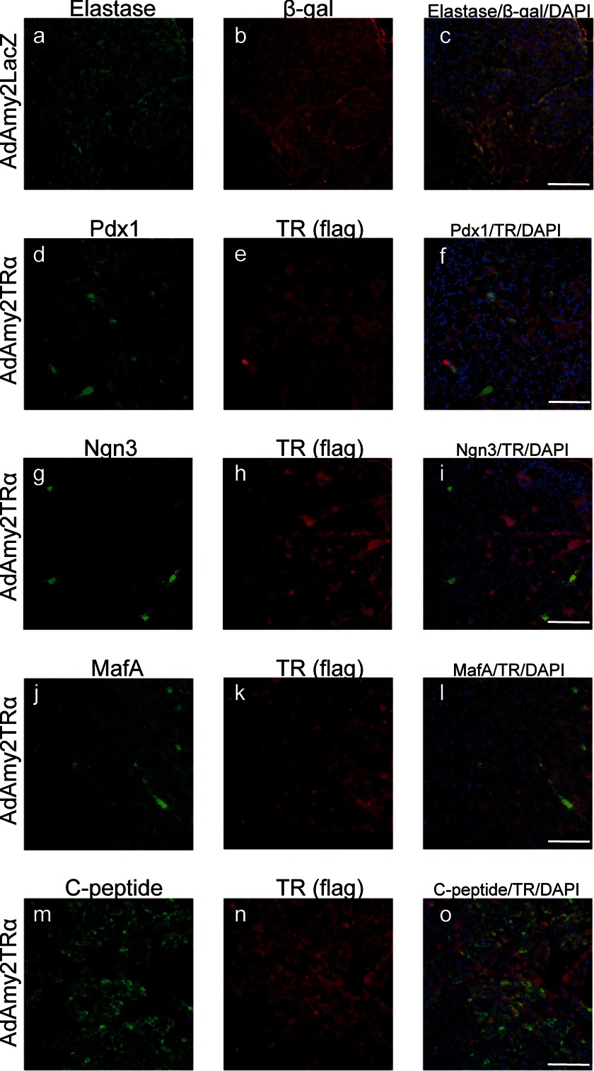
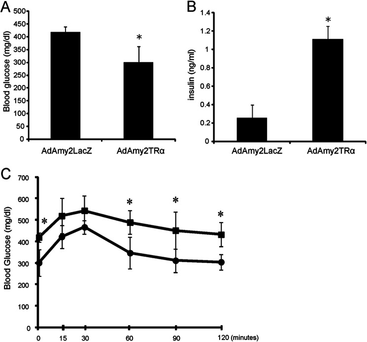
One goal of diabetic regenerative medicine is to instructively convert mature pancreatic exocrine cells into insulin-producing cells. We recently reported that ligand-bound thyroid hormone receptor α (TRα) plays a critical role in expansion of the β-cell mass during postnatal development. Here, we used an adenovirus vector that expresses TRα driven by the amylase 2 promoter (AdAmy2TRα) to induce the reprogramming of pancreatic acinar cells into insulin-producing cells. Treatment with l-3,5,3-triiodothyronine increases the association of TRα with the p85α subunit of phosphatidylinositol 3-kinase (PI3K), leading to the phosphorylation and activation of Akt and the expression of Pdx1, Ngn3, and MafA in purified acinar cells. Analyses performed with the lectin-associated cell lineage tracing system and the Cre/loxP-based direct cell lineage tracing system indicate that newly synthesized insulin-producing cells originate from elastase-expressing pancreatic acinar cells. Insulin-containing secretory granules were identified in these cells by electron microscopy. The inhibition of p85α expression by siRNA or the inhibition of PI3K by LY294002 prevents the expression of Pdx1, Ngn3, and MafA and the reprogramming to insulin-producing cells. In immunodeficient mice with streptozotocin-induced hyperglycemia, treatment with AdAmy2TRα leads to the reprogramming of pancreatic acinar cells to insulin-producing cells in vivo. Our findings suggest that ligand-bound TRα plays a critical role in β-cell regeneration during postnatal development via activation of PI3K signaling.
Keywords: Diabetes; Insulin; Pancreas; Phosphatidylinositol 3-Kinase; Thyroid Hormone.
Figures
Similar articles
-
PDX1, Neurogenin-3, and MAFA: critical transcription regulators for beta cell development and regeneration.Stem Cell Res Ther. 2017 Nov 2;8(1):240. doi: 10.1186/s13287-017-0694-z. Stem Cell Res Ther. 2017. PMID: 29096722 Free PMC article. Review.
-
Reprogramming of Pancreatic Acinar Cells to Functional Beta Cells by In Vivo Transduction of a Polycistronic Construct Containing Pdx1, Ngn3, MafA in Mice.Curr Protoc Stem Cell Biol. 2017 Feb 2;40:4A.10.1-4A.10.12. doi: 10.1002/cpsc.21. Curr Protoc Stem Cell Biol. 2017. PMID: 28152182 Free PMC article.
-
Thyroid hormones promote endocrine differentiation at expenses of exocrine tissue.Exp Cell Res. 2014 Apr 1;322(2):236-48. doi: 10.1016/j.yexcr.2014.01.030. Epub 2014 Feb 4. Exp Cell Res. 2014. PMID: 24503054
-
Protective action of liraglutide in beta cells under lipotoxic stress via PI3K/Akt/FoxO1 pathway.J Cell Biochem. 2014 Jun;115(6):1166-75. doi: 10.1002/jcb.24763. J Cell Biochem. 2014. PMID: 24415347
-
Role of MafA in pancreatic beta-cells.Adv Drug Deliv Rev. 2009 Jul 2;61(7-8):489-96. doi: 10.1016/j.addr.2008.12.015. Epub 2009 Apr 22. Adv Drug Deliv Rev. 2009. PMID: 19393272 Review.
Cited by
-
The Roles of Thyroid and Thyroid Hormone in Pancreas: Physiology and Pathology.Int J Endocrinol. 2018 Jun 14;2018:2861034. doi: 10.1155/2018/2861034. eCollection 2018. Int J Endocrinol. 2018. PMID: 30013597 Free PMC article. Review.
-
Immunohistological Study of Monkey Foveal Retina.Sci Rep. 2019 Mar 27;9(1):5258. doi: 10.1038/s41598-019-41793-y. Sci Rep. 2019. PMID: 30918305 Free PMC article.
-
Differential Gene Dosage Effects of Diabetes-Associated Gene GLIS3 in Pancreatic β Cell Differentiation and Function.Endocrinology. 2017 Jan 1;158(1):9-20. doi: 10.1210/en.2016-1541. Endocrinology. 2017. PMID: 27813676 Free PMC article.
-
Impaired oxidative endoplasmic reticulum stress response caused by deficiency of thyroid hormone receptor α.J Biol Chem. 2014 May 2;289(18):12485-93. doi: 10.1074/jbc.M113.544122. Epub 2014 Mar 18. J Biol Chem. 2014. PMID: 24644288 Free PMC article.
-
Acinar phenotype is preserved in human exocrine pancreas cells cultured at low temperature: implications for lineage-tracing of β-cell neogenesis.Biosci Rep. 2016 May 6;36(3):e00329. doi: 10.1042/BSR20150259. Print 2016 Jun. Biosci Rep. 2016. PMID: 26987985 Free PMC article.
References
-
- Edlund H. (1998) Transcribing pancreas. Diabetes 47, 1817–1823 - PubMed
-
- Schwitzgebel V. M., Scheel D. W., Conners J. R., Kalamaras J., Lee J. E., Anderson D. J., Sussel L., Johnson J. D., German M. S. (2000) Expression of neurogenin3 reveals an islet cell precursor population in the pancreas. Development 127, 3533–3542 - PubMed
-
- Murtaugh L. C., Melton D. A. (2003) Genes, signals, and lineages in pancreas development. Annu. Rev. Cell Dev. Biol. 19, 71–89 - PubMed
Publication types
MeSH terms
Substances
LinkOut - more resources
Full Text Sources
Other Literature Sources
Molecular Biology Databases
