. 2013 May 31;288(22):15771-85.
doi: 10.1074/jbc.M112.448886. Epub 2013 Apr 17.

Regulation of a dynamic interaction between two microtubule-binding proteins, EB1 and TIP150, by the mitotic p300/CBP-associated factor (PCAF) orchestrates kinetochore microtubule plasticity and chromosome stability during mitosis

Affiliations

Regulation of a dynamic interaction between two microtubule-binding proteins, EB1 and TIP150, by the mitotic p300/CBP-associated factor (PCAF) orchestrates kinetochore microtubule plasticity and chromosome stability during mitosis

Tarsha Ward et al. J Biol Chem. .

Abstract

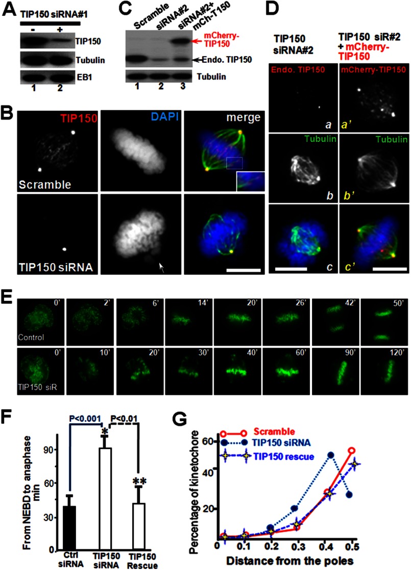
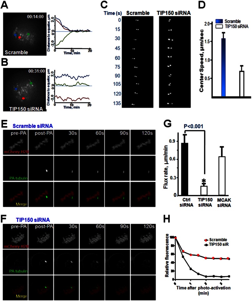
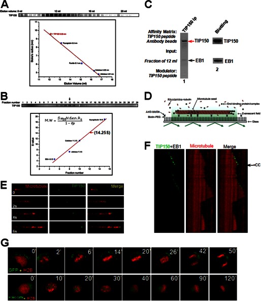
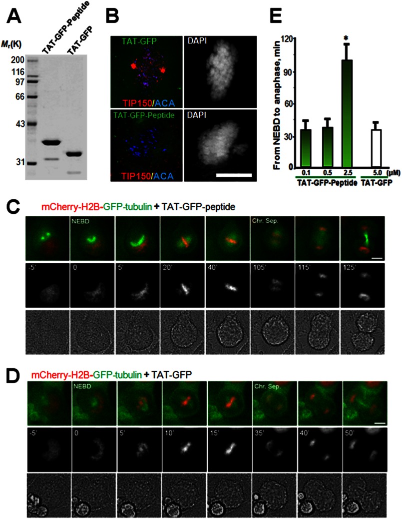
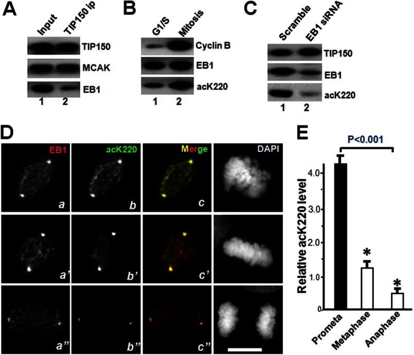
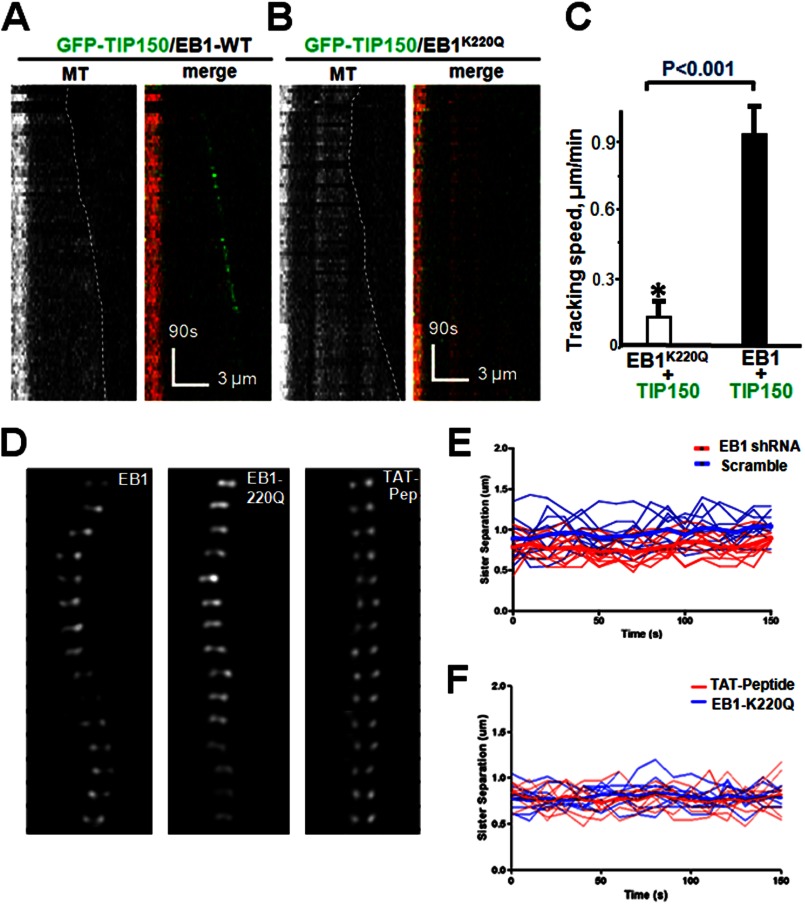
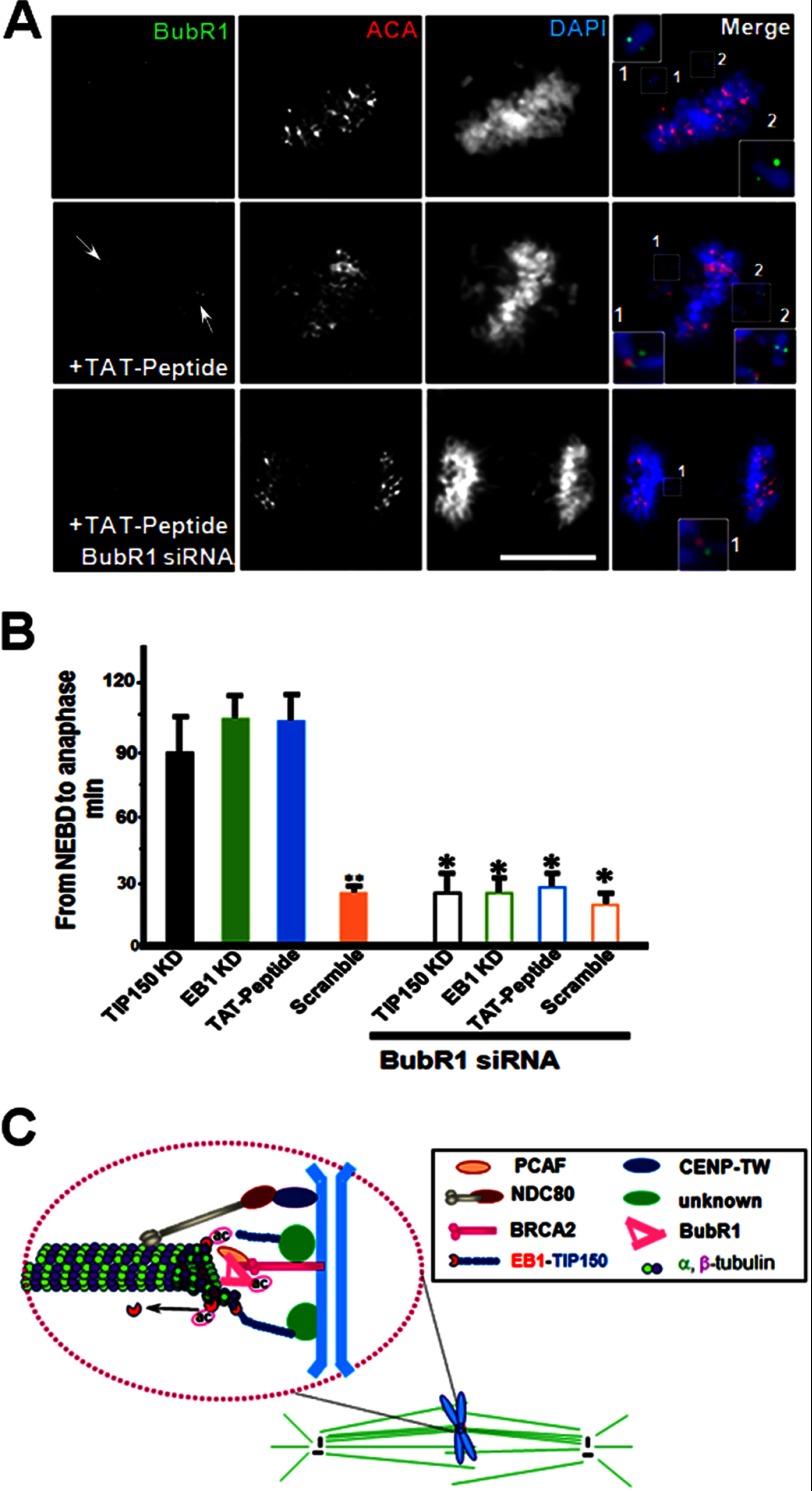
The microtubule cytoskeleton network orchestrates cellular dynamics and chromosome stability in mitosis. Although tubulin acetylation is essential for cellular plasticity, it has remained elusive how kinetochore microtubule plus-end dynamics are regulated by p300/CBP-associated factor (PCAF) acetylation in mitosis. Here, we demonstrate that the plus-end tracking protein, TIP150, regulates dynamic kinetochore-microtubule attachments by promoting the stability of spindle microtubule plus-ends. Suppression of TIP150 by siRNA results in metaphase alignment delays and perturbations in chromosome biorientation. TIP150 is a tetramer that binds an end-binding protein (EB1) dimer through the C-terminal domains, and overexpression of the C-terminal TIP150 or disruption of the TIP150-EB1 interface by a membrane-permeable peptide perturbs chromosome segregation. Acetylation of EB1-PCAF regulates the TIP150 interaction, and persistent acetylation perturbs EB1-TIP150 interaction and accurate metaphase alignment, resulting in spindle checkpoint activation. Suppression of the mitotic checkpoint serine/threonine protein kinase, BubR1, overrides mitotic arrest induced by impaired EB1-TIP150 interaction, but cells exhibit whole chromosome aneuploidy. Thus, the results identify a mechanism by which the TIP150-EB1 interaction governs kinetochore microtubule plus-end plasticity and establish that the temporal control of the TIP150-EB1 interaction by PCAF acetylation ensures chromosome stability in mitosis.

Keywords: Acetyl Coenzyme A; Cell Cycle; Genomic Instability; Mitosis; Mitotic Spindle.


Figures

FIGURE 1.
FIGURE 1.
TIP150 is essential for chromosome congression in mitosis. A, TIP150 siRNA suppresses accumulation of TIP150 protein. HeLa cells transfected with TIP150 siRNA1 and scrambled control oligonucleotides were harvested 24 h after transfection and subjected to Western blot analyses. B, knockdown of TIP150 impairs chromosome congression. HeLa cells were transfected with TIP150 siRNA or scrambled control oligonucleotides. Immunofluorescence analyses show that TIP150 exhibits a typical plus-end distribution (red) along the kinetochore microtubule (green). TIP150 siRNA treatment prevents TIP150 localization to the plus-ends and chromosome congression (bottom panel, arrow). Bar, 10 μm. C, efficiency of introduction of exogenous TIP150 in HeLa cells treated with siRNA. HeLa cells transfected with TIP150 siRNA2 targeted to 3′-UTR (lane 2), siRNA2 plus mCherry-TIP150 (lane 3), and scrambled control oligonucleotides were harvested 24 h after transfection and subjected to Western blot analyses. D, introduction of exogenously expressed TIP150 rescued the phenotype seen in TIP150-suppressed cells. HeLa cells were transfected with TIP150 siRNA2 (panels a–c) and siRNA2 plus mCherry-TIP150 (panels a′–′). Immunofluorescence analyses show that mCherry-TIP150 exhibits a typical plus-end distribution (panel a′) along the kinetochore microtubule (panel c′). Introduction of mCherry-TIP150 restored the bipolar spindle plasticity and chromosome alignment (panel c′) perturbed in siRNA2-treated cells (panel c). Bar, 10 μm. E, depletion of TIP150 perturbs chromosome alignment and mitotic progression. Real time imaging of chromosome movements in HeLa cells transfected with TIP150 siRNA and scrambled control siRNA is shown. Repression of TIP150 by siRNA resulted in chromosome misalignment and delayed cell division. F, quantitative analysis of the timing of cell division from NEBD to anaphase onset. Anaphase onset is delayed in cells treated with TIP150 siRNA (*, p < 0.01; n = 30 cells for TIP150 siRNA, Ctrl siRNA versus TIP150 siRNA; **, p < 0.01; TIP150 siRNA versus TIP150 rescue). Note that introduction of exogenously expressed TIP150 rescued the phenotype seen in TIP150 siRNA-treated cells. G, schematic illustration showing how kinetochore positions were measured by the distance from the nearest pole along the pole-pole axis and normalized for pole-pole distance as described previously (19, 28). Profiles of kinetochore position of HeLa cells transfected with scramble siRNA or TIP150 siRNA. These data were acquired from synchronized preparations as described under “Materials and Methods.” Positions were annotated in increments of 0.1. Note that introduction of exogenously expressed TIP150 rescued the phenotype seen in TIP150 siRNA-treated cells.
FIGURE 2.
FIGURE 2.
TIP150 regulates kinetochore microtubule dynamics in mitosis. A, kinetochore oscillatory dynamics was captured using time-lapse microscopy. HeLa cells were transfected with a scrambled oligonucleotide or with TIP150 siRNA (B) as described under the “Materials and Methods.” Paired sister kinetochores are color-coded to illustrate their dynamics as detailed in the text. Representative kinetochore oscillatory kymograph of scramble siRNA (A, right panel) and TIP150 siRNA-treated cells (B, right panel) are presented. C, magnified image of one kinetochore pair within a cell treated with scrambled siRNA or TIP150 siRNA (right panel). Images were acquired every 15 s as described under “Materials and Methods.” D, analysis of kinetochore velocity of scramble-transfected and TIP150-suppressed cells (n = 100 kinetochore pairs; p < 0.01). E, PA-GFP-tubulin analysis of kinetochore microtubules treated with scrambled oligonucleotide. Images display pre-photoactivated, post-activated (0 s), with 30-s intervals. F, PA-GFP-tubulin analysis of kinetochore microtubules of HeLa cells treated with TIP150 siRNA.G, quantitation of PA-GFP-tubulin pole-ward flux in TIP150-depleted HeLa cells compared with that of control scramble transfected cells. (*, p < 0.001, Ctrl siRNA versus TIP150 siRNA). H, graphical representation of GFP fluorescent decay after photoactivation from kinetochore microtubule of cells treated with TIP150 siRNA. Cells depleted of TIP150 displayed a faster decay rate compared with controls.
FIGURE 3.
FIGURE 3.
TIP150 exhibits an effective microtubule plus-end tracking activity as a tetramer. A, analysis of EB1-TIP150-purified fractions using fast protein chromatography. Constructed EB1-TIP150 complexes were subjected to gel filtration chromatography followed by SDS-PAGE fractionation. A gel stained with Coomassie Blue illustrates that the EB1-TIP150 complex eluted at 12 ml with an estimated Stokes radius of 9.55 nm. B, analysis of TIP150 complex hydrodynamics by sucrose gradient centrifugation, as described previously (29). C, biochemical characterization of TIP150-EB1 hexamer. The fraction of 12 ml from A was incubated with a TIP150 peptide antibody followed by affinity purification using a synthetic peptide (250 μm) for elution. The supernatant was then fractionated on an SDS-polyacrylamide gel followed by Coomassie Blue staining. The stoichiometry of TIP150-EB1 molecular association was calculated based on the band density of respective proteins, which is ∼4:2 (TIP150:EB1), indicating that endogenous TIP150 forms a hexameric complex with EB1. D, schematic representation of a TIRFM assay to visualize plus-end tracking activity of TIP150 using dual color tracking. E, time-lapse fluorescence images of microtubule movement with addition of 50 nm GFP-TIP150 in the presence of EB1 (50 nm). F, dual color kymographs of polymerizing microtubules with EB1 and GFP-TIP150. The histidine-TIP150(981–1368) recombinant protein (CC) was added to perturb the tetramer of TIP150 assembled in vitro (arrow). Note that the GFP-TIP150 tracks only on the growing microtubule plus-end, and addition of TIP150(981–1368) peptide removed GFP-TIP150 from the microtubule plus-end after one growth-shrinkage cycle, which resulted in an increased catastrophic frequency. G, perturbation of TIP150 tetramerization blocks chromosome alignment and mitotic progression in vivo. HeLa cells were transiently transfected to co-express GFP-TIP150(981–1368) protein and mCherry-H2B, and mitotic chromosome movements were followed by real time imaging. Overexpression of GFP-TIP150(981–1368) resulted in chromosome alignment defected and mitotic arrest, suggesting that GFP-TIP150(981–1368) acts as a dominant negative mutant.
FIGURE 4.
FIGURE 4.
Perturbation of the EB1-TIP150 interaction prevents chromosome alignment and delays anaphase onset. A, Coomassie Blue stain SDS-polyacrylamide gel was used to assess the quality and quantities of the purified TAT-GFP-His6-tagged proteins. Bacteria expressed TAT-GFP-peptide and TAT-GFP-H6 (control) are purified with nickel-nitrilotriacetic acid affinity chromatography and desalted into DMEM. Protein concentration was determined in Bradford assay. B, TAT-GFP-peptide recombinant protein disrupts EB1-TIP150 association. Aliquots of TAT-GFP or TAT-GFP-peptide (2.5 μm) was added into culture HeLa cells for 30 min followed by fixation and examination. Note that incubation of TAT-GFP-peptide librated TIP150 from kinetochore localization and resulted in a phenotype of mitotic arrest with chromosome aberrantly aligned. C and D, HeLa cells expressing mCherry-H2B and enhanced GFP-α-tubulin were synchronized with thymidine and released for 8 h to reach prometaphase. Cells were cultured in DMEM with 500 nm, 1 or 2.5 μm TAT-GFP-TIP150-d-H6 (C) or TAT-GFP-His6 (D) at 37 °C for 1–2 h before images collection. Live cell observation and imaging were performed every 5 min. Representative images are marked especially at the time of NEBD and the time of sister chromosome separation (Chr. Sep.). Bar, 10 μm. E, quantitative analysis of the timing of cell division from NEBD to anaphase onset, which is delayed in cells treated with TAT-GFP-TIP150-d (2.5 μm). The TAT-GFP-TIP150 peptide causes mitotic delay in a dose-dependent manner. (*, p < 0.001, prometa. versus metaphase or anaphase).
FIGURE 5.
FIGURE 5.
TIP150 interaction with EB1 is regulated by acetylation in mitosis. A, Western blot analysis of immunoprecipitates of TIP150, EB1, and MCAK from nocodazole-synchronized HeLa cells. EB1 proteins bind less to TIP150, relative to MCAK. B, EB1 is acetylated in mitosis. Western blot analysis of EB1 acetylation in interphase and mitotic cells. Quantitative Western blotting indicates that EB1 acetylation at Lys-220 is elevated in mitosis. C, characterization of acK220 antibody specificity. Aliquots of HeLa cells were transiently transfected to introduce siRNA and scramble control. Twenty four hours after the transfection, the HeLa cells were blocked with 100 nm nocodazole for 18 h followed by solubilization of cellular proteins. The cell lysates were fractionated by SDS-PAGE followed by Western blotting of TIP150, EB1, and acK220. The acK220 signal was abolished in EB1-suppressed cells (lane 2). D, immunofluorescence analysis showing EB1, acK220, and DAPI for DNA in mitotic HeLa cells of different stages (prometaphase, metaphase, and anaphase). Triple immunofluorescence imaging showed that EB1 acetylation level is highest at prometaphase cells (panel b), and the distribution profile of acK220 is superimposed to that of EB1 (panel c). As cells phase into metaphase and anaphase, the acK220 level decreased (panels b′ and b″). E, fluorescent intensity analysis of acetylated EB1 (acK220) in different mitotic states. EB1 has a highest level of acK220 at prometaphase cells and declines upon metaphase alignment (p < 0.001; n = 30 cells).
FIGURE 6.
FIGURE 6.
Lys-220 acetylation regulates a dynamic interaction between EB1 and TIP150. A, dual color kymographs of polymerizing microtubules with EB1 and GFP-TIP150 in vitro as illustrated in Fig. 3. B, dual color kymographs of polymerizing microtubules with acetylated EB1 and GFP-TIP150. Note that the GFP-TIP150 failed to track microtubule plus-ends in the presence of acetylated EB1K220Q. C, quantitative analysis of microtubule polymerization with EB1-GFP-TIP150 interaction compared with acetylated EB1-GFP-TIP150. Tracking speed was diminished with acetylated EB1 as determined by single-molecule tracking (*, p < 0.001; n = 30; EB1K220Q+TIP150 versus EB1-TIP150). D, perturbation of EB1 interaction by persistent Lys-220 acetylation or TIP150 peptide addition alters kinetochore oscillatory profiles in mitosis. Kinetochore oscillations of wild-type EB1 (left), K220Q (acetylated-mimicking mutant; center), and TAT-Peptide (right). Oscillations were diminished when EB1 was acetylated. E, representative kinetochore oscillatory kymograph of scramble siRNA (blue line) and EB1siRNA-treated cells (red line). F, representative kinetochore oscillatory kymograph of HeLa cells expressing persistent acetylation mimicking EB1-K220Q (blue line) or treated with TAT-peptide (red line).
FIGURE 7.
FIGURE 7.
Perturbation of EB1-TIP150 interaction activates the spindle checkpoint. A, immunofluorescence analysis shows BubR1, ACA, and DAPI for DNA. In SAC-activated cell BubR1 siRNA overrides the induced mitotic arrest by acetylated EB1-TIP150. B, quantitative analysis of the timing of cell division from NEBD to anaphase onset. Anaphase onset is delayed in cells with TIP150 knockdown, EB1 knockdown, and TAT-GFP-peptide (2.5 μm). This condition is rescued by treatment with BubR1 siRNA. **, p < 0.001, Scramble versus TP150 KD or EB1 KD or TA-peptide; *, p > 0.05, BubR1 siRNA (TIP150 KD, EB1 KD, TAT-peptide or Scramble) versus without BubR1 siRNA. C, model representation of PCAF-TIP150-EB1 interaction. In this model, PCAF is localized to kinetochore via interaction with BRCA2, and PCAF is critical for chromosome stability in mitosis (23). EB1-TIP150 interaction is required for efficient and accurate kinetochore-microtubule interactions and for achieving faithful alignment at the equator. Development of normal level of tension across bioriented kinetochore pairs, as reflected in the spatial separation of sister kinetochores, requires a temporal and dynamic regulation of EB1 acK220. The effect of temporal control of acK220 is 2-fold. First, TIP150 cooperates with EB1 to maintain a stable kinetochore-microtubule interaction, and acK220 weakens the tension across the sister kinetochores to facilitate attachment error correction. Second, the BubR1 (a checkpoint kinase that is bound to EB1/APC (adenomatous polyposis coli) and is essential for metaphase alignment (14) and disruption of EB1-TIP150 interaction) results in a prolonged activation of spindle checkpoints, as marked by BubR1 enrichment at the kinetochore, preventing activation of the APC/C (anaphase-promoting complex/cyclosome) and onset of anaphase.

References

    1. Cleveland D. W., Mao Y., Sullivan K. F. (2003) Centromeres and kinetochores: from epigenetics to mitotic checkpoint signaling. Cell 112, 407–421 - PubMed
    1. Siegel J. J., Amon A. (2012) New insights into the troubles of aneuploidy. Annu. Rev. Cell Dev. Biol. 28, 189–214 - PMC - PubMed
    1. Gordon D. J., Resio B., Pellman D. (2012) Causes and consequences of aneuploidy in cancers. Nat. Rev. Genet. 13, 189–203 - PubMed
    1. Cheeseman I. M., Desai A. (2008) Molecular architecture of the kinetochore-microtubule interface. Nat. Rev. Mol. Cell Biol. 9, 33–46 - PubMed
    1. Santaguida S., Musacchio A. (2009) The life and miracles of kinetochores. EMBO J. 28, 2511–2531 - PMC - PubMed

Publication types

MeSH terms

Substances

LinkOut - more resources