. 2013 Jul 1;305(1):C78-89.
doi: 10.1152/ajpcell.00339.2012. Epub 2013 Apr 17.

Extracellular nucleotides inhibit oxalate transport by human intestinal Caco-2-BBe cells through PKC-δ activation

Affiliations

Extracellular nucleotides inhibit oxalate transport by human intestinal Caco-2-BBe cells through PKC-δ activation

Ruhul Amin et al. Am J Physiol Cell Physiol. .

Abstract

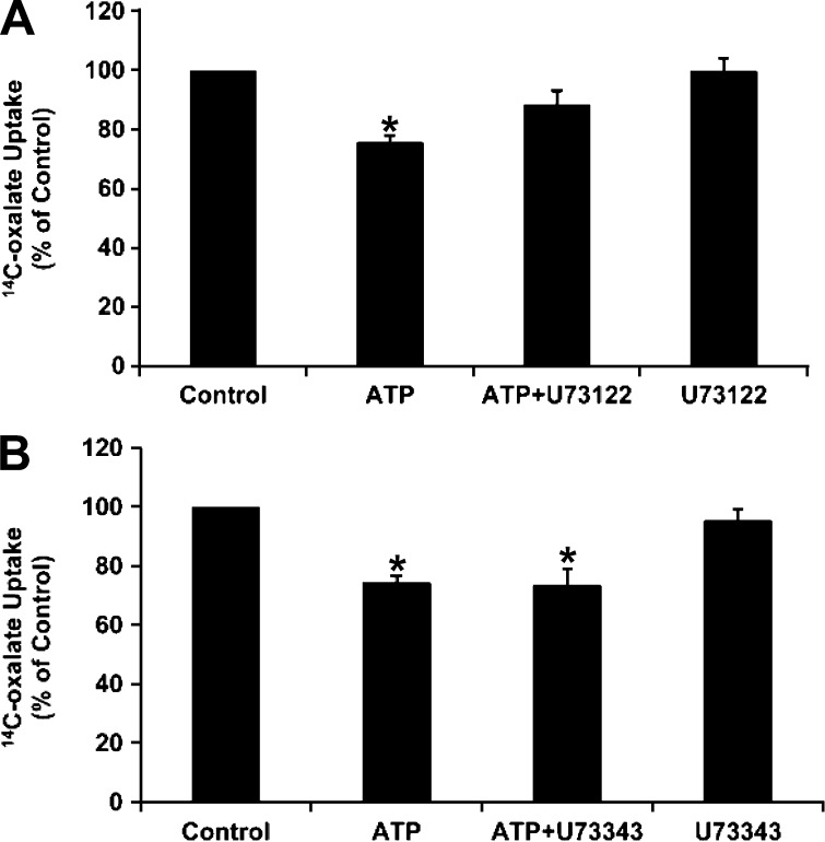
Nephrolithiasis remains a major health problem in Western countries. Seventy to 80% of kidney stones are composed of calcium oxalate, and small changes in urinary oxalate affect risk of kidney stone formation. Intestinal oxalate secretion mediated by the anion exchanger SLC26A6 plays an essential role in preventing hyperoxaluria and calcium oxalate nephrolithiasis, indicating that understanding the mechanisms regulating intestinal oxalate transport is critical for management of hyperoxaluria. Purinergic signaling modulates several intestinal processes through pathways including PKC activation, which we previously found to inhibit Slc26a6 activity in mouse duodenal tissue. We therefore examined whether purinergic stimulation with ATP and UTP affects oxalate transport by human intestinal Caco-2-BBe (C2) cells. We measured [¹⁴C]oxalate uptake in the presence of an outward Cl⁻ gradient as an assay of Cl⁻/oxalate exchange activity, ≥50% of which is mediated by SLC26A6. We found that ATP and UTP significantly inhibited oxalate transport by C2 cells, an effect blocked by the PKC inhibitor Gö-6983. Utilizing pharmacological agonists and antagonists, as well as PKC-δ knockdown studies, we observed that ATP inhibits oxalate transport through the P2Y₂ receptor, PLC, and PKC-δ. Biotinylation studies showed that ATP inhibits oxalate transport by lowering SLC26A6 surface expression. These findings are of potential relevance to pathophysiology of inflammatory bowel disease-associated hyperoxaluria, where supraphysiological levels of ATP/UTP are expected and overexpression of the P2Y₂ receptor has been reported. We conclude that ATP and UTP inhibit oxalate transport by lowering SLC26A6 surface expression in C2 cells through signaling pathways including the P2Y₂ purinergic receptor, PLC, and PKC-δ.

Keywords: P2Y2 purinergic receptor; PKC-δ; SLC26A6; phospholipase C.


Figures

Fig. 1.
Fig. 1.
Effect of ATP on [14C]oxalate uptake by Caco-2-BBE (C2) cells. A: C2 cells grown on Transwell inserts were preincubated apically with vehicle (control) or 100 μM ATP for 30 min in the culture medium, and [14C]oxalate uptake was measured. Values (means ± SE of 5 independent experiments, each done in duplicate or triplicate) were normalized to the respective control value ([14C]oxalate uptake rate = 3.95 ± 0.78 pmol·cm−2·min−1). *P < 0.003 vs. control (by 2-tailed t-test). B: C2 cells grown on plastic supports (seeded at the same time and studied concurrently as in A) were preincubated with vehicle (control) or 100 μM ATP for 30 min in the culture medium, and [14C]oxalate uptake was measured. Values (means ± SE of 4 independent experiments, each done in duplicate or triplicate) were normalized to the respective control value ([14C]oxalate uptake rate = 9.0 ± 1.08 pmol·cm−2·min−1). *P < 0.02 vs. control (by 2-tailed t-test).
Fig. 2.
Fig. 2.
Effect of UTP on [14C]oxalate uptake by C2 cells. A: C2 cells grown on Transwell inserts were preincubated apically with vehicle (control) or 100 μM UTP for 30 min in the culture medium, and [14C]oxalate uptake was measured. Values (means ± SE of 5 independent experiments, each done in duplicate or triplicate) were normalized to the respective control value ([14C]oxalate uptake rate = 2.34 ± 0.19 pmol·cm−2·min−1). *P < 0.002 vs. control (by 2-tailed t-test). B: C2 cells grown on plastic supports were preincubated with vehicle (control) or 100 μM UTP for 30 min in the culture medium, and [14C]oxalate uptake was measured. Values (means ± SE of 5 independent experiments, each done in duplicate or triplicate) were normalized to the respective control value ([14C]oxalate uptake rate = 6.84 ± 2.73 pmol·cm−2·min−1). *P < 0.0007 vs. control (by 2-tailed t-test).
Fig. 3.
Fig. 3.
Effect of ATP on [14C]oxalate uptake by T84 cells. T84 cells were preincubated with vehicle (control) or 100 μM ATP for 30 min in the culture medium, and [14C]oxalate uptake was measured. Values (means ± SE of 6 independent experiments, each done in triplicate) were normalized to the respective control value ([14C]oxalate uptake rate = 3.71 ± 1.3 pmol·cm−2·min−1). *P < 0.002 vs. control (by 2-tailed t-test).
Fig. 4.
Fig. 4.
Effect of CFTR inhibitor-172 (CFTR-172) on ATP-induced inhibition of [14C]oxalate uptake by C2 cells. C2 cells were preincubated with vehicle (control), 100 μM ATP for 30 min, 10 μM CFTR-172 for 15 min followed by 100 μM ATP for 30 min (ATP + CFTR-172), or 10 μM CFTR-172 alone for 15 min, and [14C]oxalate uptake was measured. Values (means ± SE of 4 independent experiments, each done in duplicate or triplicate) were normalized to the respective control value ([14C]oxalate uptake rate = 7.40 ± 0.93 pmol·cm−2·min−1). CFTR-172 had no significant effect on ATP-induced inhibition. B: effect of CFTR-172 on ATP-induced Cl secretion [measured as changes in short-circuit current (ΔIsc)] across C2 cells. C2 cell monolayers (seeded at the same time and studied concurrently as in A) were mounted in Ussing chambers. After a 15-min equilibration period, vehicle (control) or 10 μM CFTR-172 was added to the mucosal side of matched pairs of monolayers for 15 min; then ATP (100 μM) was added to the mucosal side to elicit Cl secretion. Values (means ± SE of 14 monolayers per group) are presented as peak Isc elicited by ATP. CFTR-172 significantly inhibited ATP-induced Cl secretion (ΔIsc). *P < 0.001 vs. control (by ANOVA).
Fig. 5.
Fig. 5.
Effect of P2Y1 and P2Y2 receptor agonists on [14C]oxalate uptake by C2 cells. C2 cells were preincubated with vehicle (control), 100 μM ATP, 100 μM UTP, 1 μM 2-thiouridine-5′-triphosphate (2-thio-UTP, a P2Y2 agonist), or 1 μM MRS-2365 (a P2Y1 agonist) for 30 min in the culture medium, and [14C]oxalate uptake was measured. Values (means ± SE of 5–8 independent experiments, each done in duplicate or triplicate) were normalized to the respective control value ([14C]oxalate uptake rate = 3.66 ± 0.41 pmol·cm−2·min−1). *P < 0.001 vs. control (by ANOVA).
Fig. 6.
Fig. 6.
Effect of the PLC inhibitor U-73122 and its inactive analog U-73343 on ATP-induced inhibition of [14C]oxalate uptake by C2 cells. A: C2 cells were preincubated with vehicle (control), 100 μM ATP for 30 min, 10 μM U-73122 for 30 min followed by 100 μM ATP with continued presence of U-73122 (ATP + U-73122) for 30 min, or 10 μM U-73122 alone for 60 min, and [14C]oxalate uptake was measured. Values (means ± SE of 8 independent experiments, each done in duplicate or triplicate) were normalized to the respective control value ([14C]oxalate uptake rate = 8.48 ± 1.55 pmol·cm−2·min−1). *P < 0.001 and P < 0.05 vs. control and ATP + U-73122, respectively (by ANOVA). B: C2 cells were preincubated with vehicle (control), 100 μM ATP for 30 min, 10 μM U-73343 for 30 min followed by 100 μM ATP with continued presence of U-73343 (ATP + U-73343) for 30 min, or 10 μM U-73343 alone for 60 min, and [14C]oxalate uptake was measured. Values (means ± SE of 7 independent experiments, each done in duplicate or triplicate) were normalized to the respective control value ([14C]oxalate uptake rate = 6.20 ± 0.73 pmol·cm−2·min−1). U-73343 had no significant effect on inhibition induced by ATP. *P < 0.001 vs. control (by ANOVA).
Fig. 7.
Fig. 7.
Effect of the PKC inhibitor Gö-6983 on UTP-induced inhibition of [14C]oxalate uptake by C2 cells. C2 cells were preincubated with vehicle (control) or 100 μM UTP for 30 min in the culture medium, and [14C]oxalate uptake was measured. [14C]oxalate uptake was also measured in the presence of Gö-6983 (1 μM) for 30 min followed by 100 μM UTP with continued presence of Gö-6983 (UTP + Gö-6983) or 1 μM Gö-6983 alone for 60 min. Values (means ± SE of 6 independent experiments, each done in duplicate or triplicate) were normalized to the respective control value ([14C]oxalate uptake rate = 2.61 ± 0.50 pmol·cm−2·min−1). Gö-6983 completely and significantly reduced the inhibition induced by UTP: *P < 0.01 and P < 0.05 vs. control and UTP + Gö-6983, respectively (by ANOVA).
Fig. 8.
Fig. 8.
PKC-δ knockdown in C2 cells using small interfering RNA (siRNA). A: representative Western blot of PKC-δ protein expression in C2 cell lysate (30 μg protein/lane). UT, untransfected cells; CON, C2 cells transfected with negative control siRNA; PKC-δ, C2 cells transfected with siRNA targeting PKC-δ. Anti-β-actin antibody was used to normalize loading of protein in each lane. B: densitometry of immunoblot results. Western blot band density was quantified using ImageJ software. Values (means ± SE for 4 independent experiments) represent PKC-δ abundance relative to β-actin and are presented as percentage of UT value. *P < 0.001 and P < 0.01 vs. UT and CON, respectively (by ANOVA). C: effect of PKC-δ knockdown on ATP-induced inhibition of [14C]oxalate uptake by C2 cells. C2 cells transfected with negative control siRNA or PKC-δ siRNA were preincubated with vehicle (control) or 100 μM ATP [(control siRNA + ATP) or (PKC-δ siRNA + ATP)] for 30 min in the culture medium, and [14C]oxalate uptake was measured. Values (means ± SE of 6 independent experiments, each done in duplicate or triplicate) were normalized to the respective control value ([14C]oxalate uptake rate = 9.13 ± 0.73 pmol·cm−2·min−1). *P < 0.01 and P < 0.05 vs. control and PKC-δ siRNA + ATP, respectively (by ANOVA).
Fig. 9.
Fig. 9.
Effect of ATP on SLC26A6 (A6) protein expression. A: representative Western blot of total and surface biotinylated SLC26A6. C2 cells were preincubated with vehicle (CON) or 100 μM ATP for 30 min, and SLC26A6 protein expression was evaluated in cell lysate [total (50 μg protein/lane)] and after streptavidin precipitation of surface biotinylated proteins from 2,000 μg of initial cell lysate (surface). Anti-GAPDH antibody was used to verify equal loading of protein in each lane. B and C: densitometry of immunoblot results. Western blot band density was quantified using ImageJ software. Values (means ± SE for 5 independent experiments for cells grown on plastic supports and 6 independent experiments for cells grown on Transwell inserts) represent total SLC26A6 abundance relative to GAPDH and surface biotinylated SLC26A6 relative to total SLC26A6 and are presented as a percentage of the respective control value. *P < 0.003 vs. CON (by paired t-test).

References

    1. Alexander RT, Hemmelgarn BR, Wiebe N, Bello A, Morgan C, Samuel S, Klarenbach SW, Curhan GC, Tonelli M, Alberta Kidney Disease Network Kidney stones and kidney function loss: a cohort study. Br Med J 345: e5287, 2012 - PMC - PubMed
    1. Ao M, Venkatasubramanian J, Boonkaewwan C, Ganesan N, Syed A, Benya RV, Rao MC. Lubiprostone activates Cl− secretion via cAMP signaling and increases membrane CFTR in the human colon carcinoma cell line, T84. Dig Dis Sci 56: 339– 351, 2011 - PubMed
    1. Arpin M, Blair L, Coudrier E, Dudouet B, Finidori J, Carcia A, Huet C, Pringault E, Robine S, Sahuguillo-Merino C, et al. Villin, a specific marker for some epithelia specialized in transport, to study the differentiation of intestinal and kidney cells in vivo and in a human colon adenocarcinoma line HT29 in culture. Mol Aspects Med 10: 257– 272, 1988 - PubMed
    1. Asplin JR. Hyperoxaluric calcium nephrolithiasis. Endocrinol Metab Clin North Am 31: 927– 949, 2002 - PubMed
    1. Athman R, Louvard D, Robine S. The epithelial cell cytoskeleton and intracellular trafficking. III. How is villin involved in the actin cytoskeleton dynamics in intestinal cells? Am J Physiol Gastrointest Liver Physiol 283: G496– G502, 2002 - PubMed

Publication types

MeSH terms