

Sex differences in the neural correlates of affective experience
- PMID: 23596188
- PMCID: PMC4014103
- DOI: 10.1093/scan/nst030
Sex differences in the neural correlates of affective experience
Abstract
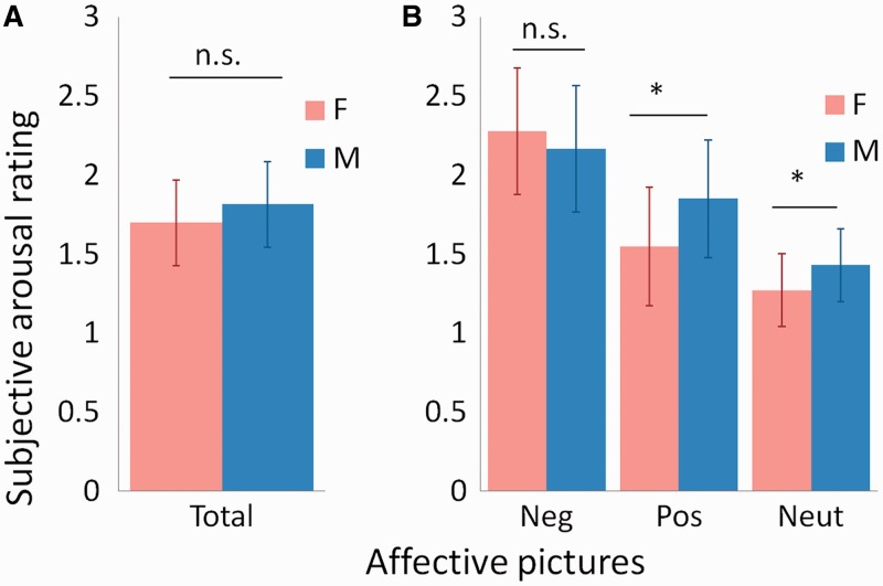
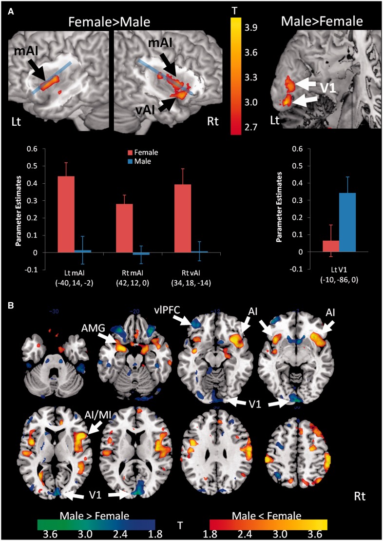
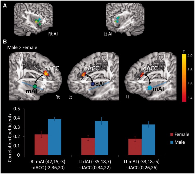
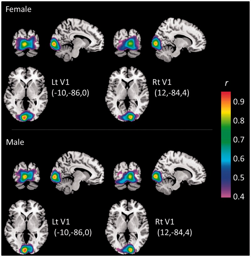
People believe that women are more emotionally intense than men, but the scientific evidence is equivocal. In this study, we tested the novel hypothesis that men and women differ in the neural correlates of affective experience, rather than in the intensity of neural activity, with women being more internally (interoceptively) focused and men being more externally (visually) focused. Adult men (n = 17) and women (n = 17) completed a functional magnetic resonance imaging study while viewing affectively potent images and rating their moment-to-moment feelings of subjective arousal. We found that men and women do not differ overall in their intensity of moment-to-moment affective experiences when viewing evocative images, but instead, as predicted, women showed a greater association between the momentary arousal ratings and neural responses in the anterior insula cortex, which represents bodily sensations, whereas men showed stronger correlations between their momentary arousal ratings and neural responses in the visual cortex. Men also showed enhanced functional connectivity between the dorsal anterior insula cortex and the dorsal anterior cingulate cortex, which constitutes the circuitry involved with regulating shifts of attention to the world. These results demonstrate that the same affective experience is realized differently in different people, such that women's feelings are relatively more self-focused, whereas men's feelings are relatively more world-focused.
Keywords: anterior cingulate; anterior insula; extraceptive; fMRI; interoceptive.
Figures
Similar articles
-
How Hot Are They? Neural Correlates of Genital Arousal: An Infrared Thermographic and Functional Magnetic Resonance Imaging Study of Sexual Arousal in Men and Women.J Sex Med. 2018 Feb;15(2):217-229. doi: 10.1016/j.jsxm.2017.12.006. Epub 2018 Jan 6. J Sex Med. 2018. PMID: 29310889
-
Brain structures mediating cardiovascular arousal and interoceptive awareness.Brain Res. 2007 Apr 13;1141:178-87. doi: 10.1016/j.brainres.2007.01.026. Epub 2007 Jan 12. Brain Res. 2007. PMID: 17296169
-
Subgenual anterior cingulate-insula resting-state connectivity as a neural correlate to trait and state stress resilience.Brain Cogn. 2018 Jul;124:73-81. doi: 10.1016/j.bandc.2018.05.001. Epub 2018 May 11. Brain Cogn. 2018. PMID: 29758439
-
Neural substrates of sexual arousal are not sex dependent.Proc Natl Acad Sci U S A. 2019 Jul 30;116(31):15671-15676. doi: 10.1073/pnas.1904975116. Epub 2019 Jul 15. Proc Natl Acad Sci U S A. 2019. PMID: 31308220 Free PMC article.
-
[Identifying distinct components in the cerebral treatment of visual sexual information through functional neuroimaging].J Soc Biol. 2004;198(3):247-53. J Soc Biol. 2004. PMID: 15662942 Review. French.
Cited by
-
Affective and cognitive brain-networks are differently integrated in women and men while experiencing compassion.Front Psychol. 2022 Sep 13;13:992935. doi: 10.3389/fpsyg.2022.992935. eCollection 2022. Front Psychol. 2022. PMID: 36176793 Free PMC article.
-
Early adverse life events are associated with altered brain network architecture in a sex- dependent manner.Neurobiol Stress. 2017 Feb 17;7:16-26. doi: 10.1016/j.ynstr.2017.02.003. eCollection 2017 Dec. Neurobiol Stress. 2017. PMID: 28239631 Free PMC article.
-
Human threat circuits: Threats of pain, aggressive conspecific, and predator elicit distinct BOLD activations in the amygdala and hypothalamus.Front Psychiatry. 2023 Jan 9;13:1063238. doi: 10.3389/fpsyt.2022.1063238. eCollection 2022. Front Psychiatry. 2023. PMID: 36733415 Free PMC article.
-
Social reappraisal of emotions is linked with the social presence effect in the default mode network.Front Psychiatry. 2023 Mar 23;14:1128916. doi: 10.3389/fpsyt.2023.1128916. eCollection 2023. Front Psychiatry. 2023. PMID: 37032933 Free PMC article.
-
The most important attribute: stakeholder perspectives on what matters most in a physician.Curr Oncol. 2020 Apr;27(2):100-106. doi: 10.3747/co.27.5489. Epub 2020 May 1. Curr Oncol. 2020. PMID: 32489252 Free PMC article.
References
-
- Aikins DE, Anticevic A, Kiehl KA, Krystal JH. Sex-related differences in amygdala activity influences immediate memory. Neuroreport. 2010;21(4):273–76. doi:10.1097/WNR.0b013e328335b3f9. - PubMed
-
- Allman J, Hakeem A, Watson K. Two phylogenetic specializations in the human brain. Neuroscientist. 2002;8(4):335–46. - PubMed
-
- Ansfield ME. Smiling when distressed: when a smile is a frown turned upside down. Personality and Social Psychology Bulletin. 2007;33(6):763–75. doi:10.1177/0146167206297398. - PubMed
-
- Augustine JR. Circuitry and functional aspects of the insular lobe in primates including humans. Brain Research Brain Research Reviews. 1996;22(3):229–44. - PubMed
-
- Baron-Cohen S. The Essential Difference: The Truth About the Male and Female Brain. New York: Basic Books; 2003.
Publication types
MeSH terms
Grants and funding
- P41-RR14075/RR/NCRR NIH HHS/United States
- DP1OD003312/OD/NIH HHS/United States
- S10 RR019307/RR/NCRR NIH HHS/United States
- P41 RR014075/RR/NCRR NIH HHS/United States
- K02 AG029480/AG/NIA NIH HHS/United States
- 1S10RR023401/RR/NCRR NIH HHS/United States
- DP1 OD003312/OD/NIH HHS/United States
- S10 RR023043/RR/NCRR NIH HHS/United States
- S10 RR023401/RR/NCRR NIH HHS/United States
- R01 AG030311/AG/NIA NIH HHS/United States
- R01-AG029480/AG/NIA NIH HHS/United States
- 1S10RR019307/RR/NCRR NIH HHS/United States
- 1S10RR023043/RR/NCRR NIH HHS/United States
LinkOut - more resources
Full Text Sources
Other Literature Sources
Molecular Biology Databases
