. 2013 Apr 30;110(18):7136-41.
doi: 10.1073/pnas.1302378110. Epub 2013 Apr 17.

Molecular view of an electron transfer process essential for iron-sulfur protein biogenesis

Affiliations

Molecular view of an electron transfer process essential for iron-sulfur protein biogenesis

Lucia Banci et al. Proc Natl Acad Sci U S A. .

Abstract

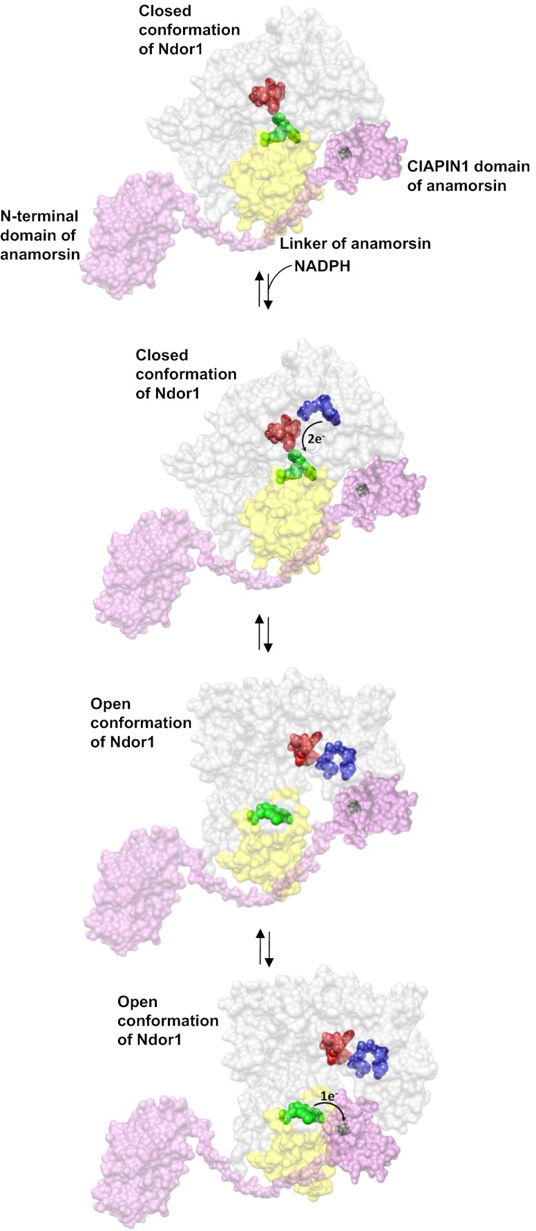
Biogenesis of iron-sulfur cluster proteins is a highly regulated process that requires complex protein machineries. In the cytosolic iron-sulfur protein assembly machinery, two human key proteins--NADPH-dependent diflavin oxidoreductase 1 (Ndor1) and anamorsin--form a stable complex in vivo that was proposed to provide electrons for assembling cytosolic iron-sulfur cluster proteins. The Ndor1-anamorsin interaction was also suggested to be implicated in the regulation of cell survival/death mechanisms. In the present work we unravel the molecular basis of recognition between Ndor1 and anamorsin and of the electron transfer process. This is based on the structural characterization of the two partner proteins, the investigation of the electron transfer process, and the identification of those protein regions involved in complex formation and those involved in electron transfer. We found that an unstructured region of anamorsin is essential for the formation of a specific and stable protein complex with Ndor1, whereas the C-terminal region of anamorsin, containing the [2Fe-2S] redox center, transiently interacts through complementary charged residues with the FMN-binding site region of Ndor1 to perform electron transfer. Our results propose a molecular model of the electron transfer process that is crucial for understanding the functional role of this interaction in human cells.


Conflict of interest statement

The authors declare no conflict of interest.

Figures

Fig. 1.
Fig. 1.
Structure of the FMN-binding domain of Ndor1. (A) Ribbon representation of the crystal structure of the FMN-binding domain of Ndor1. FMN is shown as beige spheres. (B) Superimposition of the crystal structures of the FMN-binding domains of Ndor1 (green, FMN in beige spheres) and of the human NADPH-cytochrome P450 reductase (PDB ID: 1B1C) (red, FMN in orange spheres). The regions with structural variations between the two proteins are shown in gray. (C) Molecular surface of the FMN-binding domain of Ndor1 (Left) and of the human NADPH-cytochrome P450 reductase (Right) colored according to their electrostatic potential. The views are equivalent in terms of the orientation of the protein backbone. The colors of the molecular surface indicate positive (blue), neutral (white), and negative (red) electrostatic potentials. The FMN molecule is shown as orange spheres.
Fig. 2.
Fig. 2.
Structural characterization of [2Fe-2S]-CIAPIN1-single. (A) 1H-15N IR-HSQC-AP spectrum, optimized to detect fast relaxing 1H resonances, showing 13 backbone NH resonances of the [2Fe-2S]-CIAPIN1-single, 10 of which (in black) were completely lost in the standard diamagnetic 1H-15N HSQC experiment (the three residues also detected in the diamagnetic 1H-15N HSQC are shown in gray. (B) Representative model of the CIAPIN1 domain of anamorsin derived from a molecular dynamics simulation of 100 ns. The [2Fe-2S] cluster bound to the CX8CX2CXC motif is shown; the four cysteines coordinating the [2Fe-2S] cluster and the four cysteines of the CX2CX7CX2C motif are shown.
Fig. 3.
Fig. 3.
Protein–protein interaction between the FMN-binding domain of Ndor1 and [2Fe-2S]-anamorsin as characterized by NMR. (A) TROSY 1H-15N HSQC at 308 K, acquired at 900 MHz, of the 15N-labeled oxidized FMN domain (construct 1–174 aa) before (red) and after (black) the addition of 1 eq of unlabeled oxidized [2Fe-2S]-anamorsin. (B) Overlay of 1H-15N HSQC spectra of 15N-labeled oxidized FMN-binding domain at 298 K, acquired at 800 MHz, before (black) and after (red) addition of 1 eq of [2Fe-2S]-CIAPIN1-single. (Inset) Chemical shift variations of selected FMN-binding domain residues upon addition of increasing amounts of [2Fe-2S]-CIAPIN1-single (0%, 50%, and 100%).
Fig. 4.
Fig. 4.
Mapping the binding interface of the FMN-binding domain of Ndor1 interacting with [2Fe-2S]-anamorsin. (A and B) Ribbon representations of the FMN-binding domain showing as spheres backbone NHs of the residues experiencing chemical shift variations upon interaction with (A) [2Fe-2S]-anamorsin and (B) [2Fe-2S]-CIAPIN1-single. Large blue spheres, residues showing large chemical shift changes and characterized by relative solvent accessibility above 50%; small blue spheres, residues showing large chemical shift changes but featuring relative solvent accessibility lower than 50%; large cyan spheres, residues showing small chemical shift changes with concomitant broadening effects and with relative solvent accessibility above 50%; small cyan spheres, residues showing small chemical shift changes with concomitant broadening effects and with relative solvent accessibility lower than 50%. Positively charged (gray), negatively charged (red), and hydrophobic (yellow) side chains which are highly solvent exposed in the interacting region are indicated.
Fig. 5.
Fig. 5.
Model of the electron transfer process between Ndor1 and anamorsin. Anamorsin (in pink) can be tightly bound to both closed and open conformational states of Ndor1 (in gray) due to the specific recognition between an unstructured region of anamorsin and a region of the FMN-binding domain that is solvent exposed in both open and closed conformations (in yellow). The N-terminal domain of anamorsin is not involved in the protein–protein recognition process. Upon NADPH (in blue) binding, electrons can efficiently be transferred in the closed conformation of Ndor1 to FAD (in red) and then to FMN (in green) to produce the hydroquinone state of FMN. Interflavin electron transfer in Ndor1 significantly populates the open conformation, which exposes to the solvent the FMN moiety and allows the formation of a transient interaction with the [2Fe-2S] cluster region of anamorsin located in the CIAPIN1 domain. The latter interaction allows an efficient transfer of one electron from the hydroquinone state of FMN to the [2Fe-2S] cluster (in black).

References

    1. Johnson DC, Dean DR, Smith AD, Johnson MK. Structure, function, and formation of biological iron-sulfur clusters. Annu Rev Biochem. 2005;74:247–281. - PubMed
    1. Rouault TA, Tong WH. Iron-sulfur cluster biogenesis and human disease. Trends Genet. 2008;24(8):398–407. - PMC - PubMed
    1. Lill R. Function and biogenesis of iron-sulphur proteins. Nature. 2009;460(7257):831–838. - PubMed
    1. Meyer J. Iron-sulfur protein folds, iron-sulfur chemistry, and evolution. J Biol Inorg Chem. 2008;13(2):157–170. - PubMed
    1. Qi W, Cowan JA. Structural, mechanistic and coordination chemistry of relevance to the biosynthesis of iron-sulfur and related iron cofactors. Coord Chem Rev. 2011;255(7–8):688–699. - PMC - PubMed

Publication types

MeSH terms

Associated data

LinkOut - more resources