

Divergent kinetics of proliferating T cell subsets in simian immunodeficiency virus (SIV) infection: SIV eliminates the "first responder" CD4+ T cells in primary infection
- PMID: 23596288
- PMCID: PMC3676080
- DOI: 10.1128/JVI.00027-13
Divergent kinetics of proliferating T cell subsets in simian immunodeficiency virus (SIV) infection: SIV eliminates the "first responder" CD4+ T cells in primary infection
Abstract
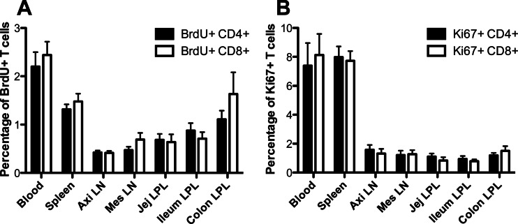
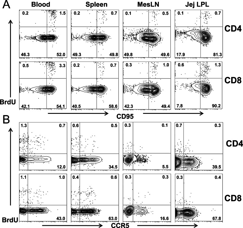
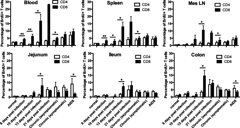
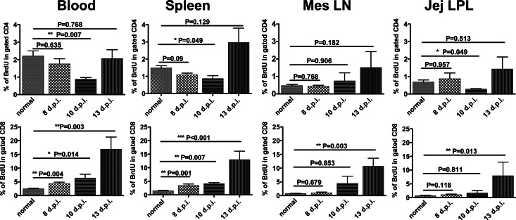
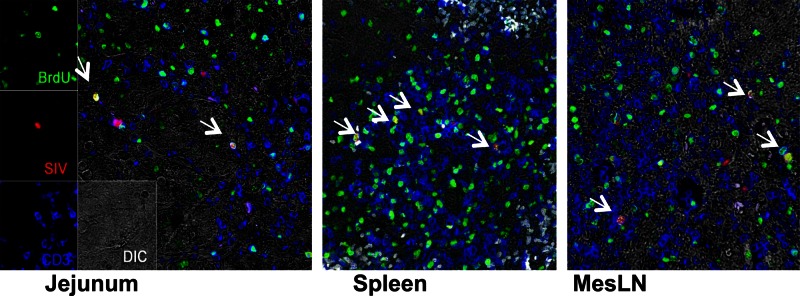
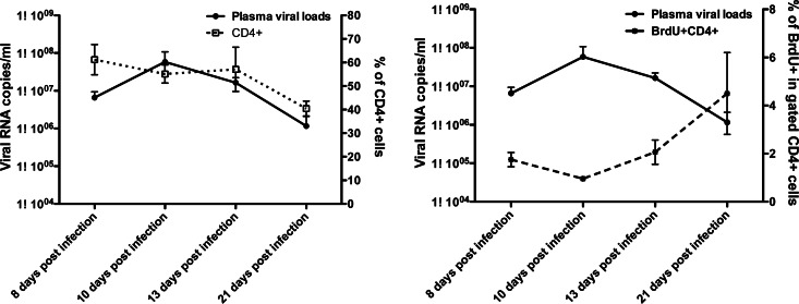
Although increased lymphocyte turnover in chronic human immunodeficiency virus and simian immunodeficiency virus (SIV) infection has been reported in blood, there is little information on cell turnover in tissues, particularly in primary SIV infection. Here we examined the levels of proliferating T cell subsets in mucosal and peripheral lymphoid tissues of adult macaques throughout SIV infection. To specifically label cells in S-phase division, all animals were inoculated with bromodeoxyuridine 24 h prior to sampling. In healthy macaques, the highest levels of proliferating CD4(+) and CD8(+) T cells were in blood and, to a lesser extent, in spleen. Substantial percentages of proliferating cells were also found in intestinal tissues, including the jejunum, ileum, and colon, but very few proliferating cells were detected in lymph nodes (axillary and mesenteric). Moreover, essentially all proliferating T cells in uninfected animals coexpressed CD95 and many coexpressed CCR5 in the tissues examined. Confocal microscopy also demonstrated that proliferating cells were substantial viral target cells for SIV infection and viral replication. After acute SIV infection, percentages of proliferating CD4(+) and CD8(+) T cells were significantly higher in tissues of chronically infected macaques and macaques with AIDS than in those of the controls. Surprisingly, however, we found that proliferating CD4(+) T cells were selectively decreased in very early infection (8 to 10 days postinoculation [dpi]). In contrast, levels of proliferating CD8(+) T cells rapidly increased after SIV infection, peaked by 13 to 21 dpi, and thereafter remained significantly higher than those in the controls. Taken together, these findings suggest that SIV selectively infects and destroys dividing, nonspecific CD4(+) T cells in acute infection, resulting in homeostatic changes and perhaps continuing loss of replication capacity to respond to nonspecific and, later, SIV-specific antigens.
Figures
References
-
- Chase A, Zhou Y, Siliciano RF. 2006. HIV-1-induced depletion of CD4+ T cells in the gut: mechanism and therapeutic implications. Trends Pharmacol. Sci. 27:4–7 - PubMed
-
- Picker LJ, Hagen SI, Lum R, Reed-Inderbitzin EF, Daly LM, Sylwester AW, Walker JM, Siess DC, Piatak M, Jr, Wang C, Allison DB, Maino VC, Lifson JD, Kodama T, Axthelm MK. 2004. Insufficient production and tissue delivery of CD4+ memory T cells in rapidly progressive simian immunodeficiency virus infection. J. Exp. Med. 200:1299–1314 - PMC - PubMed
-
- Hellerstein MK, McCune JM. 1997. T cell turnover in HIV-1 disease. Immunity 7:583–589 - PubMed
-
- Hellerstein MK. 1999. Measurement of T-cell kinetics: recent methodologic advances. Immunol. Today 20:438–441 - PubMed
Publication types
MeSH terms
Grants and funding
LinkOut - more resources
Full Text Sources
Other Literature Sources
Research Materials
