. 2013 Jun;87(12):7176-84.
doi: 10.1128/JVI.03533-12. Epub 2013 Apr 17.

Sodium taurocholate cotransporting polypeptide mediates woolly monkey hepatitis B virus infection of Tupaia hepatocytes

Affiliations

Sodium taurocholate cotransporting polypeptide mediates woolly monkey hepatitis B virus infection of Tupaia hepatocytes

Guocai Zhong et al. J Virol. 2013 Jun.

Abstract

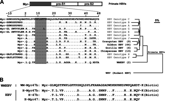
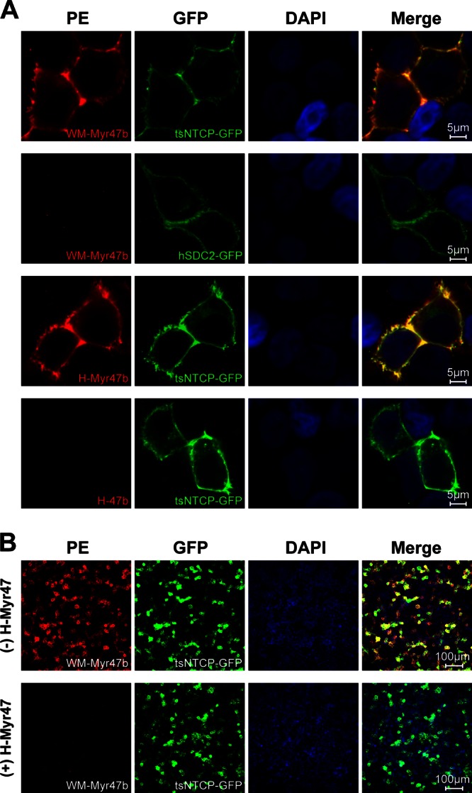
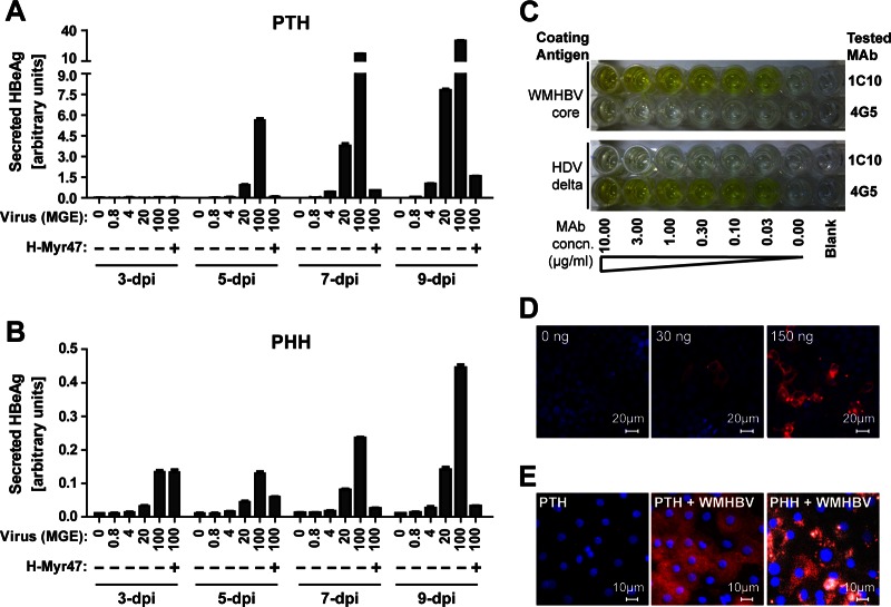
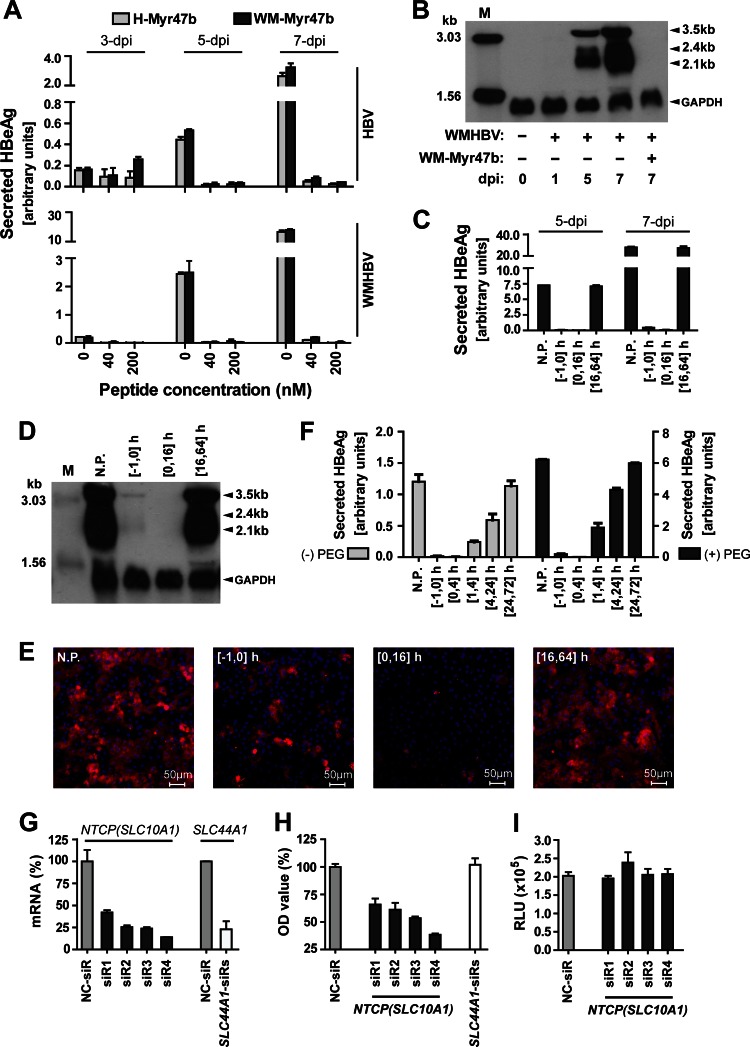
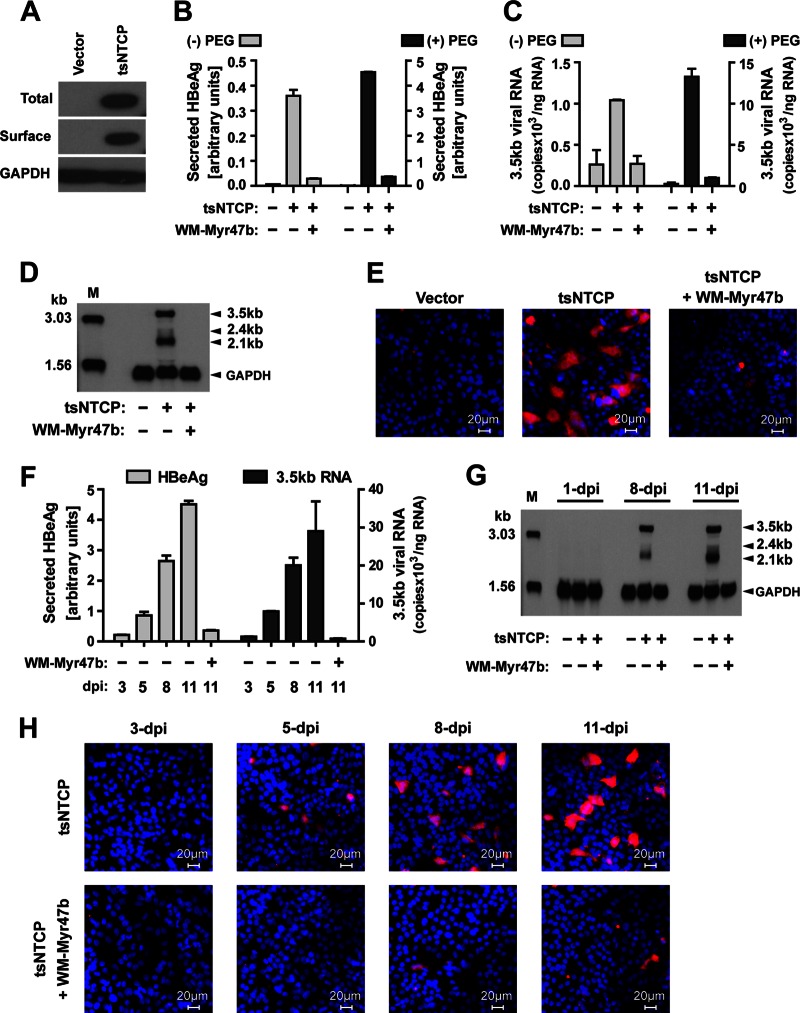
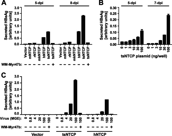
Primary Tupaia hepatocytes (PTHs) are susceptible to woolly monkey hepatitis B virus (WMHBV) infection, but the identity of the cellular receptor(s) mediating WMHBV infection of PTHs remains unclear. Recently, sodium taurocholate cotransporting polypeptide (NTCP) was identified as a functional receptor for human hepatitis B virus (HBV) infection of primary human and Tupaia hepatocytes. In this study, a synthetic pre-S1 peptide from WMHBV was found to bind specifically to cells expressing Tupaia NTCP (tsNTCP) and it efficiently blocked WMHBV entry into PTHs; silencing of tsNTCP in PTHs significantly inhibited WMHBV infection. Ectopic expression of tsNTCP rendered HepG2 cells susceptible to WMHBV infection. These data demonstrate that tsNTCP is a functional receptor for WMHBV infection of PTHs. The result also indicates that NTCP's orthologs likely act as a common cellular receptor for all known primate hepadnaviruses.


Figures

Fig 1
Fig 1
Sequence alignment of the pre-S1 N-terminal domain of primate hepadnaviruses and phylogenetic analysis of the L proteins. (A) Amino acid sequences of the receptor-binding region (aa −10 to 48 or 2 to 48) in the pre-S1 domain of human HBVs were aligned with the corresponding regions of other primate HBVs. The residue numbering was based on HBV genotype D. The GenBank accession numbers of the hepadnaviruses compared herein are as follows: HBV genotype A, JQ687533; genotype B, JX978431; genotype C, AY167095; genotype D, X02496; genotype E, GQ161835; genotype F, DQ899142; genotype G, AP007264; genotype H, AB205010; genotype J, AB486012; chimpanzee HBV, AF222322; gorilla HBV, AJ131567; orangutan HBV, EU155825; gibbon HBV, U46935; WMHBV, AY226578. For nonhuman primate HBVs, the values in parentheses are the numbers of recorded complete genomes of the corresponding viruses. The receptor-binding motif of human HBV (aa 9 to 15) and the corresponding regions of other primate HBVs are shaded in gray. The phylogenetic tree was based on the amino acid sequences of the L proteins of the compared primate HBVs and woodchuck hepatitis virus (GenBank accession number J02442), a rodent hepadnavirus. The distance along the horizontal axis among isolates is proportional to the amino acid divergence. The scale bar (upper right) represents 5% divergence. (B) Amino acid sequences of four synthetic peptides used in this study. The peptide WM-Myr47b corresponds to aa 2 to 47 of the L protein of WMHBV (GenBank accession number AY226578). The amino acid sequences of the three HBV peptides are derived from aa 2 to 47 of the L protein of the genotype C isolate (GenBank accession number AY167095). Peptides of this region were synthesized with or without a lysine residue at the C terminus for biotinylation and with or without myristoylation modification at the N terminus. Myr, myristoyl group.
Fig 2
Fig 2
Specific binding of WM-Myr47b peptide to cell surface tsNTCP. (A) 293T cells were transiently transfected with plasmids encoding tsNTCP-GFP or hSDC2-GFP as a control. Transfected cells were cultured in PMM for 36 to 48 h, blocked with 3% bovine serum albumin–phosphate-buffered saline for 1 h, and then incubated with the indicated peptides at 400 nM at 37°C for 2 h. Subsequently, the cells were fixed with 4% paraformaldehyde, stained with 0.6 μg/ml PE-streptavidin, and visualized with a Zeiss LSM 510 Meta confocal microscope. (B) 293T cells transfected with tsNTCP-GFP were blocked with 3% bovine serum albumin–phosphate-buffered saline in the absence (top) or presence (bottom) of 800 nM nonbiotinylated HBV pre-S1 peptide, H-Myr47, followed by washing and incubation with the biotinylated WMHBV pre-S1 peptide, WM-Myr47b, at 400 nM at 37°C for 2 h. Cells were then stained and visualized as described for panel A. DAPI, 4′,6-diamidino-2-phenylindole.
Fig 3
Fig 3
Validation of the ELISA kit for WMHBV HBeAg detection and MAb 1C10 for WMHBV HBcAg detection. (A, B) PTHs or PHHs were inoculated for 16 h with the indicated MGE of WMHBV in the presence of 4% PEG 8000 with or without 200 nM H-Myr47 peptide. Cell culture supernatants were collected every 2 days. Supernatants from different days postinoculation were measured by ELISA. The relative quantity of HBeAg is presented as arbitrary units per 50 μl of culture supernatant, which was calculated by multiplying the optical density at 450 nm by the dilution factors. (C) 1C10 is a mouse MAb developed by the hybridoma technique with recombinant WMHBV core protein and recognizes the core proteins of both WMHBV and HBV. Protein G-purified MAb 1C10 and control MAb 4G5 targeting HDV delta antigen were serially diluted in 3% bovine serum albumin–phosphate-buffered saline, incubated in an ELISA plate precoated with 5 μg/ml recombinant WMHBV core protein or HDV delta antigen, and detected by goat anti-mouse IgG-horseradish peroxidase conjugate. 1C10 recognized WMHBV core protein (yellow wells, top) but not HDV delta antigen. (D) Huh7 cells in a 48-well plate were transfected with 0, 30, or 150 ng of WMHBV core expression plasmid. At 24 h after transfection, cells were fixed, permeabilized, and stained with 5 μg/ml MAb 1C10. (E) PTHs or PHHs infected with WMHBV at an MGE of 0 or 100 in panels A and B were fixed at 14 dpi, and intracellular HBcAg of WMHBV was stained with 5 μg/ml of MAb 1C10. Red, WMHBV HBcAg; blue, cell nucleus; white, autofluorescence.
Fig 4
Fig 4
Inhibition of WMHBV infection in PTHs by tsNTCP-binding peptide or tsNTCP-specific siRNAs. (A) Inhibition of HBV and WMHBV infection by peptide WM-Myr47b or H-Myr47b. PTHs were inoculated with Huh7-produced WMHBV at an MGE of 100 or HBV (genotype D) at an MGE of 200 in the presence of 4% PEG 8000 for 16 h. The indicated peptide was added immediately before inoculation and was present during the inoculation process. Cell culture supernatants were collected every 2 days, and secreted HBeAg was measured by ELISA. The relative quantity of HBeAg is presented as arbitrary units per 50 μl of culture supernatant, which was calculated by multiplying the optical density at 450 nm by the dilution factors. (B) PTHs in six-well plates were inoculated with WMHBV as in panel A. At 0, 1, 5, and 7 dpi, cells were homogenized for total RNA extraction. A 10-μg sample of total RNA was separated on a 1.5% agarose gel and then blotted to Hybond-N+ positively charged nylon membrane (GE Healthcare Life Sciences) by overnight capillary transfer in 10× SSC (1× SSC is 0.15 M NaCl plus 0.015 M sodium citrate). Hybridization was performed at 52°C for 6 h with a random primed digoxigenin-dUTP (Roche Applied Science)-labeled probe covering the whole genome of WMHBV and a probe spanning human glyceraldehyde 3-phosphate dehydrogenase (GAPDH) transcript variant 1 mRNA nucleotides 181 to 690. Hybridized probes were detected with anti-digoxigenin antibody–alkaline phosphatase conjugate and CSPD chemiluminescence substrate (Roche Applied Science). The chemiluminescence signal was detected with BioMax Light chemiluminescence film (Sigma). Samples (100 pg) of denatured 3,032- and 1,554-bp PCR fragments of the WMHBV genome were included as markers. (C to E) Blocking of WMHBV infection by adding WM-Myr47b at different time points. PTHs were inoculated with WMHBV at an MGE of 100 in the presence of 4% PEG 8000 for 16 h. The WM-Myr47b peptide was added to PTHs at 200 nM and incubated for different times as indicated. The time of virus addition to cells is designated 0 h. N.P., no peptide treatment. (C) Cell culture supernatants were collected every 2 days, and secreted HBeAg was measured on the indicated days by ELISA. (D) At 7 dpi, infected PTHs in a six-well plate were homogenized for total RNA extraction and 10 μg of RNA was subjected to Northern blot analysis as in panel B. Lane M corresponds to 10 pg of denatured 3,032- and 1,554-bp PCR fragments from the WMHBV genome. (E) At 14 dpi, infected PTHs were fixed with 4% paraformaldehyde and intracellular HBcAg of WMHBV was stained with 5 μg/ml MAb 1C10 and then with Alexa 488-conjugated goat anti-mouse IgG (Life Technologies). Fluorescence images were captured with a Zeiss LSM 510 Meta confocal microscope. Red, WMHBV HBcAg; blue, cell nucleus. (F) WM-Myr47b-mediated inhibition of WMHBV infection at 4 h after inoculation. PTHs were inoculated with WMHBV at an MGE of 100 in the presence or absence of 4% PEG 8000 for 4 h. The WM-Myr47b peptide was added at 200 nM to PTHs and incubated for the times indicated. The time at which the virus was added to the cells was marked as 0 h. Secreted HBeAg was measured by ELISA at 5 dpi. N.P., no peptide treatment. (G) Validation of the knockdown efficiencies of siRNAs at the mRNA level. Freshly isolated PTHs were transfected with 20 nM siRNA targeting tsNTCP (SLC10A1: siR1, siR2, siR3, and siR4), or tsSLC44A1 (SLC44A1-siRs) or with a nontargeting control siRNA (NC-siR). Three days after transfection, PTHs were lysed for total RNA isolation and cDNA preparation. Tupaia NTCP (SLC10A1) and SLC44A1 mRNA levels were then determined by qRT-PCR with tsNTCP- and tsSLC44A1-specific primers, respectively. qRT-PCR was performed with the SYBR Premix Ex Taq kit (TaKaRa, Tokyo, Japan) on an ABI Fast 7500 RT-PCR system (Applied Biosystems). Data are presented as the relative mRNA levels of NTCP and SLC44A1 compared to that in cells transfected with the negative-control siRNA (NC-siR). (H) WMHBV infection of PTHs is reduced by tsNTCP knockdown. Freshly isolated PTHs were transfected as described for panel G. Three days after transfection, cells were inoculated with WMHBV at an MGE of 100 for 16 h. The culture medium was refreshed every 2 days, and secreted HBeAg was measured at 5 dpi. Data are presented as the relative optical density (OD) value compared to the optical density at 450 nm for HBeAg from cells transfected with the negative-control siRNA (NC-siR). (I) Infection with control virus lenti-VSV-G was not affected by tsNTCP knockdown. Freshly isolated PTHs were transfected as described for panel G. Three days after transfection, cells were inoculated with lenti-VSV-G for 16 h. The culture medium was refreshed every 2 days, and cells were lysed for luciferase activity detection at 5 dpi. RLU, relative luminescence units.
Fig 5
Fig 5
Tupaia NTCP expression supports WMHBV infection of HepG2 cells. (A) Western blot analysis of tsNTCP expression in HepG2 cells. HepG2 cells were transfected with a plasmid encoding C9-tagged tsNTCP or a vector control plasmid. Six hours after transfection, the culture medium was changed to PMM and the cells were cultured for an additional 24 h. The cells were subjected to Western blot analysis for the levels of total and surface expression of tsNTCP protein as previously described (6). (B, C) HepG2 cells were transfected with a plasmid encoding tsNTCP or a vector control plasmid. Six hours after transfection, the culture medium was changed to PMM and the cells were cultured for an additional 24 h. Subsequently, the cells were inoculated with WMHBV at an MGE of 100 in the absence or presence of 4% PEG 8000 and with or without 200 nM peptide WM-Myr47b. (B) The culture medium was refreshed every 2 to 3 days, and secreted HBeAg was measured at 11 dpi. (C) The cells were then lysed for total RNA preparation and quantification of the 3.5-kb WMHBV precore RNA/pgRNA by qRT-PCR with the SYBR Premix Ex Taq kit and primers targeting the viral core gene. The levels of 3.5-kb viral RNA are presented as copy numbers of viral RNA per nanogram of total cellular RNA. (D, E) HepG2 cells were transfected as described for panel B and inoculated with WMHBV in the presence of 4% PEG 8000 with or without 200 nM peptide WM-Myr47b. At 11 dpi, the cells were either examined by Northern blot analysis with 15 μg of total RNA (D) or immunostained for intracellular HBcAg (E). (F to H) Kinetics of WMHBV infection of tsNTCP-complemented HepG2 cells. HepG2 cells were transfected as described for panel A and inoculated with WMHBV in the presence of 4% PEG 8000 with or without 200 nM peptide WM-Myr47b. Levels of secreted HBeAg (F), viral RNAs (F and G), and intracellular HBcAg (H) were determined by ELISA, qRT-PCR, Northern blot analysis, and intracellular immunostaining, respectively, on the days indicated. Lane M in panels D and G corresponds to 100 pg of denatured 3,032- and 1,554-bp PCR fragments from the WMHBV genome. In panels E and H, red is WMHBV HBcAg and blue is the cell nucleus.
Fig 6
Fig 6
Species specificity of NTCP-mediated WMHBV infection and correlation of WMHBV infection with the amount of tsNTCP and the virus inoculation dose. (A) Species specificity of NTCP-mediated WMHBV infection of HepG2 cells. HepG2 cells were transfected with plasmids encoding NTCPs of different species or with a vector control plasmid. The cells were then cultured in PMM for 24 h. Subsequently, the cells were inoculated with WMHBV in the presence of 4% PEG 8000 with or without 200 nM WM-Myr47b peptide. Secreted HBeAg was determined by ELISA on the days indicated. (B) Correlation of the amount of transfected tsNTCP plasmid and the WMHBV infection level. tsNTCP plasmid was serially diluted as indicated, and the total amount of DNA was compensated with the vector plasmid to ensure that the same amount of DNA was transfected into HepG2 cells in 48-well plates. The transfected cells were then cultured and inoculated as described for panel A. Secreted HBeAg was determined by ELISA on the days indicated. (C) Correlation of the virus inoculation dose and the WMHBV infection level. HepG2 cells were transfected with a plasmid encoding tsNTCP or hNTCP or with a vector control plasmid. The cells were then cultured in PMM for 24 h. Subsequently, the cells were inoculated for 16 h with WMHBV at the indicated MGE in the presence of 4% PEG 8000 with or without 200 nM WM-Myr47b peptide. Secreted HBeAg in the culture supernatant was measured by ELISA at 11 dpi.

References

    1. Ott JJ, Stevens GA, Groeger J, Wiersma ST. 2012. Global epidemiology of hepatitis B virus infection: new estimates of age-specific HBsAg seroprevalence and endemicity. Vaccine 30:2212–2219 - PubMed
    1. Hagenbuch B, Meier PJ. 1994. Molecular cloning, chromosomal localization, and functional characterization of a human liver Na+/bile acid cotransporter. J. Clin. Invest. 93:1326–1331 - PMC - PubMed
    1. Stieger B. 2011. The role of the sodium-taurocholate cotransporting polypeptide (NTCP) and of the bile salt export pump (BSEP) in physiology and pathophysiology of bile formation. Handb. Exp. Pharmacol. 201:205–259 - PubMed
    1. Hu NJ, Iwata S, Cameron AD, Drew D. 2011. Crystal structure of a bacterial homologue of the bile acid sodium symporter ASBT. Nature 478:408–411 - PMC - PubMed
    1. Mareninova O, Shin JM, Vagin O, Turdikulova S, Hallen S, Sachs G. 2005. Topography of the membrane domain of the liver Na+-dependent bile acid transporter. Biochemistry 44:13702–13712 - PubMed

Publication types

MeSH terms

Substances