Suppressed Th17 levels correlate with elevated PIAS3, SHP2, and SOCS3 expression in CD4 T cells during acute simian immunodeficiency virus infection
- PMID: 23596301
- PMCID: PMC3676085
- DOI: 10.1128/JVI.00600-13
Suppressed Th17 levels correlate with elevated PIAS3, SHP2, and SOCS3 expression in CD4 T cells during acute simian immunodeficiency virus infection
Abstract
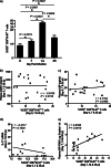
T helper 17 (Th17) cells play an important role in mucosal immune homeostasis and maintaining the integrity of the mucosal epithelial barrier. Loss of Th17 cells has been extensively documented during human immunodeficiency virus (HIV) and simian immunodeficiency virus (SIV) infections. The lack of effective repopulation of Th17 cells has been associated with chronic immune activation mediated by the translocation of microbial products. Using ex vivo analysis of purified peripheral blood CD4 T cells from SIV-infected rhesus macaques, we show that the suppression of interleukin-17 (IL-17) expression correlated with upregulated expression of negative regulatory genes PIAS3, SHP2, and SOCS3 in CD4 T cells. Suppressed Th17 expression was accompanied by elevated levels of soluble CD14 (sCD14) and lipopolysaccharide binding protein (LBP) in the plasma during early stages of infection. Plasma viral loads rather than sCD14 or LBP levels correlated with acute immune activation. Additionally, we observed a significant increase in the expression of CD14 on peripheral blood monocytes that correlated with IL-23 expression and markers of microbial translocation. Taken together, our results provide new insights into the early events associated with acute SIV pathogenesis and suggest additional mechanisms playing a role in suppression of Th17 cells.
Figures
Similar articles
-
Mucosal T Helper 17 and T Regulatory Cell Homeostasis Correlate with Acute Simian Immunodeficiency Virus Viremia and Responsiveness to Antiretroviral Therapy in Macaques.AIDS Res Hum Retroviruses. 2019 Mar;35(3):295-305. doi: 10.1089/AID.2018.0184. Epub 2019 Jan 2. AIDS Res Hum Retroviruses. 2019. PMID: 30398361 Free PMC article.
-
IL-21 Therapy Controls Immune Activation and Maintains Antiviral CD8+ T Cell Responses in Acute Simian Immunodeficiency Virus Infection.AIDS Res Hum Retroviruses. 2017 Nov;33(S1):S81-S92. doi: 10.1089/aid.2017.0160. AIDS Res Hum Retroviruses. 2017. PMID: 29140110 Free PMC article.
-
Differential Dynamics of Regulatory T-Cell and Th17 Cell Balance in Mesenteric Lymph Nodes and Blood following Early Antiretroviral Initiation during Acute Simian Immunodeficiency Virus Infection.J Virol. 2019 Sep 12;93(19):e00371-19. doi: 10.1128/JVI.00371-19. Print 2019 Oct 1. J Virol. 2019. PMID: 31315987 Free PMC article.
-
Th17 cells, HIV and the gut mucosal barrier.Curr Opin HIV AIDS. 2010 Mar;5(2):173-8. doi: 10.1097/COH.0b013e328335eda3. Curr Opin HIV AIDS. 2010. PMID: 20543596 Review.
-
Th17 cells in pathogenic simian immunodeficiency virus infection of macaques.Curr Opin HIV AIDS. 2010 Mar;5(2):141-5. doi: 10.1097/COH.0b013e32833653ec. Curr Opin HIV AIDS. 2010. PMID: 20543591 Free PMC article. Review.
Cited by
-
Oncostatin M Suppresses Activation of IL-17/Th17 via SOCS3 Regulation in CD4+ T Cells.J Immunol. 2017 Feb 15;198(4):1484-1491. doi: 10.4049/jimmunol.1502314. Epub 2017 Jan 16. J Immunol. 2017. PMID: 28093521 Free PMC article.
-
Microbial Translocation and Inflammation Occur in Hyperacute Immunodeficiency Virus Infection and Compromise Host Control of Virus Replication.PLoS Pathog. 2016 Dec 7;12(12):e1006048. doi: 10.1371/journal.ppat.1006048. eCollection 2016 Dec. PLoS Pathog. 2016. PMID: 27926931 Free PMC article.
-
Suppression of transforming growth factor β receptor 2 and Smad5 is associated with high levels of microRNA miR-155 in the oral mucosa during chronic simian immunodeficiency virus infection.J Virol. 2015 Mar;89(5):2972-8. doi: 10.1128/JVI.03248-14. Epub 2014 Dec 24. J Virol. 2015. PMID: 25540365 Free PMC article.
-
Loss and dysregulation of Th17 cells during HIV infection.Clin Dev Immunol. 2013;2013:852418. doi: 10.1155/2013/852418. Epub 2013 May 23. Clin Dev Immunol. 2013. PMID: 23762098 Free PMC article. Review.
-
Inflammation status in HIV-positive individuals correlates with changes in bone tissue quality after initiation of ART.J Antimicrob Chemother. 2019 May 1;74(5):1381-1388. doi: 10.1093/jac/dkz014. J Antimicrob Chemother. 2019. PMID: 30768163 Free PMC article.
References
-
- Douek DC, Picker LJ, Koup RA. 2003. T cell dynamics in HIV-1 infection. Annu. Rev. Immunol. 21:265–304 - PubMed
-
- Brenchley JM, Price DA, Schacker TW, Asher TE, Silvestri G, Rao S, Kazzaz Z, Bornstein E, Lambotte O, Altmann D, Blazar BR, Rodriguez B, Teixeira-Johnson L, Landay A, Martin JN, Hecht FM, Picker LJ, Lederman MM, Deeks SG, Douek DC. 2006. Microbial translocation is a cause of systemic immune activation in chronic HIV infection. Nat. Med. 12:1365–1371 - PubMed
-
- Cecchinato V, Trindade CJ, Laurence A, Heraud JM, Brenchley JM, Ferrari MG, Zaffiri L, Tryniszewska E, Tsai WP, Vaccari M, Parks RW, Venzon D, Douek DC, O'Shea JJ, Franchini G. 2008. Altered balance between Th17 and Th1 cells at mucosal sites predicts AIDS progression in simian immunodeficiency virus-infected macaques. Mucosal Immunol. 1:279–288 - PMC - PubMed
-
- Favre D, Lederer S, Kanwar B, Ma ZM, Proll S, Kasakow Z, Mold J, Swainson L, Barbour JD, Baskin CR, Palermo R, Pandrea I, Miller CJ, Katze MG, McCune JM. 2009. Critical loss of the balance between Th17 and T regulatory cell populations in pathogenic SIV infection. PLoS Pathog. 5:e1000295 doi:10.1371/journal.ppat.1000295 - DOI - PMC - PubMed
-
- Raffatellu M, Santos RL, Verhoeven DE, George MD, Wilson RP, Winter SE, Godinez I, Sankaran S, Paixao TA, Gordon MA, Kolls JK, Dandekar S, Baumler AJ. 2008. Simian immunodeficiency virus-induced mucosal interleukin-17 deficiency promotes Salmonella dissemination from the gut. Nat. Med. 14:421–428 - PMC - PubMed
Publication types
MeSH terms
Substances
Grants and funding
LinkOut - more resources
Full Text Sources
Other Literature Sources
Research Materials
Miscellaneous

