. 2013 May 15;190(10):5237-46.
doi: 10.4049/jimmunol.1203017. Epub 2013 Apr 17.

IL-17 stimulates differentiation of human anti-inflammatory macrophages and phagocytosis of apoptotic neutrophils in response to IL-10 and glucocorticoids

Affiliations

IL-17 stimulates differentiation of human anti-inflammatory macrophages and phagocytosis of apoptotic neutrophils in response to IL-10 and glucocorticoids

Gaetano Zizzo et al. J Immunol. .

Abstract

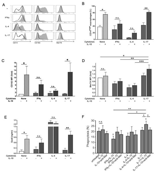
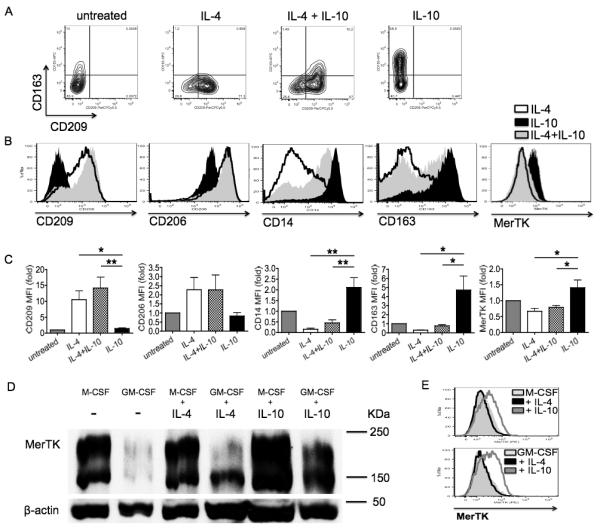
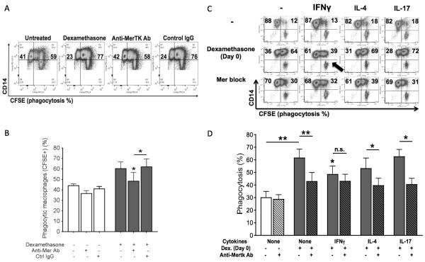
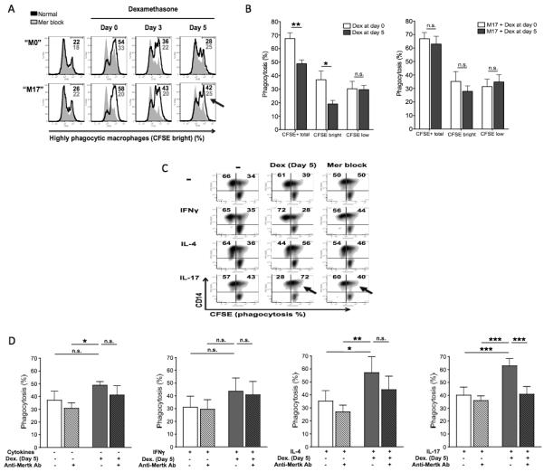
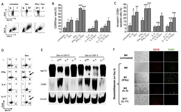
Exposure of human monocytes/macrophages to anti-inflammatory agents, such as IL-10 or glucocorticoids, can lead to two separate fates: either Fas/CD95-mediated apoptosis or differentiation into regulatory and efferocytic M2c (CD14(bright)CD16(+)CD163(+)Mer tyrosine kinase(+)) macrophages. We found that the prevalent effect depends on the type of Th cytokine environment and on the stage of monocyte-to-macrophage differentiation. In particular, the presence of IFN-γ (Th1 inflammation) or the prolonged exposure to IL-4 (chronic Th2 inflammation) promotes apoptosis of monocytes/macrophages and causes resistance to M2c differentiation, thus provoking impaired clearance of apoptotic neutrophils, uncontrolled accumulation of apoptotic cells, and persistent inflammation. In contrast, the presence of IL-17 (Th17 environment) prevents monocyte/macrophage apoptosis and elicits intense M2c differentiation, thus ensuring efficient clearance of apoptotic neutrophils and restoration of anti-inflammatory conditions. Additionally, the Th environment affects the expression of two distinct Mer tyrosine kinase isoforms: IL-4 downregulates the membrane isoform but induces an intracellular and Gas6-dependent isoform, whereas IFN-γ downregulates both and IL-17 upregulates both. Our data support an unexpected role for IL-17 in orchestrating resolution of innate inflammation, whereas IFN-γ and IL-4 emerge as major determinants of IL-10 and glucocorticoid resistance.


Figures

Figure 1
Figure 1. Macrophage response to IL-10: IL-17, but not IFNγ or IL-4, elicits M2c differentiation and MerTK-dependent efferocytosis
(A) Human monocytes were sorted from healthy PBMCs through negative selection magnetic beads and cultured in complete medium without cytokines (M0 differentiation), or in the presence of IFNγ (10 ng/ml; M1 differentiation), IL-4 (20 ng/ml; M2a differentiation) or IL-17 (100 ng/ml; M17 differentiation). On day 5, cells were treated with IL-10 (50 ng/ml) for an additional 3 days (M2c polarization). Grey histograms, cells cultured with IL-10; white histograms, cells cultured without IL-10. Expression of the M2c markers CD14, CD163 and MerTK was measured by flow cytometry. (B-E) To measure autocrine production of Gas6 along with surface expression of M2c markers, cells were cultured for 4 days in serum-free medium with IFNγ, IL-4 or IL-17, in the presence or absence of IL-10. Expression levels of CD14 (B), CD163 (C), and MerTK (D) were assessed by flow cytometry and depicted as percentages of CD14bright cells or mean fluorescence intensity (MFI) fold variation compared to levels obtained with culturing cells without IL-10. Gas6 levels (E) in culture supernatants were quantified by ELISA. Data are representative of four independent experiments. (F) Early ANs were obtained by incubating healthy human neutrophils in 10% FBS-RPMI for 20 hours. CFSE-labeled ANs were added for 30 minutes, at a 5:1 ratio, to CD14-labeled macrophages differentiated for 7-8 days in complete medium without cytokines or in the presence of IFNγ, IL-4 or IL-17, with or without IL-10 in the last 3 days. For inhibition studies, macrophages were pre-incubated with a goat polyclonal anti-human MerTK antibody (5 μg/ml; R&D Systems) for 30 minutes before the addition of ANs. Data are representative of four independent experiments. Pooled data are represented as mean values ± SEM. *P <0.05; **P <0.01; ***P <0.001; n.s., not significant.
Figure 2
Figure 2. IL-4 impedes IL-10 differentiation of M2c macrophages, but induces Gas6 and a 150-KDa intracellular MerTK isoform
(A-C) Macrophages were differentiated for 8 days in complete medium plus IL-4 (20 ng/ml; M2a differentiation), IL-10 (50 ng/ml; M2c differentiation), or both cytokines from day 3 for 5 days. Expression levels of CD163, CD209, CD206, CD14 and MerTK was assessed by flow cytometry, and depicted in (C) as MFI fold variation compared to levels obtained with culturing cells without any cytokine. (D-E) To increase the number of terminally differentiated macrophages on day 8 and clearly detect a minor MerTK isoform, macrophages were differentiated in the presence of growth factors M-CSF (50 ng/ml) or GM-CSF (100 ng/ml) from day 0, adding IL-4 or IL-10 on day 3 for an additional 5 days. A 150-KDa (intracellular) MerTK isoform was observed by Western blot. The 200-KDa (surface) MerTK isoform was detected by both Western blot (D) and flow cytometry (E). Data are representative of three independent experiments. Pooled data are represented as mean values ± SEM. *P <0.05; **P <0.01.
Figure 3
Figure 3. Early response to glucocorticoids: IL-17 and IL-4, but not IFNγ, allow M2c differentiation and MerTK/Gas6 induction
Monocytes were cultured for 4 days in serum-free medium with or without dexamethasone from day 0 (100 nM; M2c differentiation), in the presence or absence of IFNγ (10 ng/ml), IL-4 (20 ng/ml) or IL-17 (100 ng/ml). Expression levels of CD14 (A), CD16 (B), MerTK (C) were measured by flow cytometry and depicted as MFI fold variation compared to untreated cells. Gas6 levels (D) in culture supernatants were measured by ELISA. Expression of 200-KDa and 150-KDa MerTK isoforms (E) was assessed by Western blot. Data are representative of four independent experiments. Pooled data are represented as mean values ± SEM. *P <0.05; **P <0.01; ***P <0.001.
Figure 4
Figure 4. Early response to glucocorticoids: IL-17 and IL-4, but not IFNγ, allow differentiation of MerTK-dependent efferocytic macrophages
CFSE-labeled ANs were added for 30 minutes, at a 5:1 ratio, to CD14-labeled macrophages differentiated for 7-8 days in complete medium with or without dexamethasone from day 0 (100 nM; M2c differentiation), in the presence of absence of IFNγ (10 ng/ml), IL-4 (20 ng/ml) or IL-17 (100 ng/ml). For inhibition studies, macrophages were pre-incubated with a goat polyclonal anti-human MerTK antibody (5 μg/ml; R&D Systems) for 30 minutes before the addition of ANs. Specificity of anti-MerTK antibody was preliminarily assessed by comparing its effects with those exerted by a goat polyclonal control IgG (5 μg/ml; SouthernBiotech). All data shown are representative of four independent experiments. Pooled data are represented as mean values ± SEM. *P <0.05; **P <0.01; n.s., not significant.
Figure 5
Figure 5. Late response to glucocorticoids: IL-17, but not IFNγ or IL-4, promotes MerTK-dependent efferocytosis by terminally differentiated macrophages
(A-B) Macrophages were differentiated for 8 days in complete medium in the presence or absence of IL-17 (100 ng/ml). Dexamethasone was added from day 0 (M2c differentiation) or during monocyte-to macrophage differentiation, from day 3 or from day 5. CFSE-labeled ANs were then coincubated with CD14-labeled macrophages, at a 5:1 ratio, for 30 minutes. Among macrophages, in (A) we could distinguish non-efferocytic CFSEnull macrophages (left peak), moderately efferocytic CFSElow macrophages (central peak) and highly efferocytic CFSEbright macrophages (right peak). Numerical values refer to percentages of CFSEbright macrophages. Grey peaks refer to assays performed in the presence of a goat polyclonal anti-human MerTK blocking antibody (5 μg/ml; R&D Systems), added for 30 minutes prior to macrophage coincubation with ANs. CFSEtotal macrophages in (B) refers to total efferocytic (CFSEbright plus CFSElow) macrophages. (C-D) Cells were cultured in complete medium with no cytokines (M0 differentiation), or in the presence of IFNγ (10 ng/ml; M1 differentiation), IL-4 (20 ng/ml; M2a differentiation) or IL-17 (100 ng/ml; M17 differentiation); from day 5, cells were treated with dexamethasone (100 nM) for an additional 3 days (M2c polarization). CFSE-labeled ANs were then coincubated with CD14-labeled macrophages, at a 5:1 ratio, for 30 minutes. For inhibition studies, macrophages were pre-incubated with a goat polyclonal anti-human MerTK antibody (5 μg/ml; R&D Systems) for 30 minutes before the addition of ANs. Data are representative of four independent experiments. Pooled data are represented as mean values ± SEM. *P <0.05; **P <0.01; ***P <0.001; n.s., not significant.
Figure 6
Figure 6. Response to IL-10 and glucocorticoids: IL-17, but not IFNγ o r IL-4, constantly protects macrophages from CD95 upregulation and apoptosis
(A) Cells were cultured for 4 days in serum-free medium with or without dexamethasone from day 0 (100 nM; M2c differentiation), in the presence or absence of IFNγ (10 ng/ml). Panels representing CD16 and MerTK expression, along with panels representing Side Scatter (SSC) and Forward Scattor (FSC) are shown. Data shown are representative of four independent experiments. (B-D) Cells were cultured for 3 days in complete medium in the presence or absence of IFNγ (10 ng/ml; M1 differentiation), IL-4 (20 ng/ml; M2a differentiation) or IL-17 (100 ng/ml; M17 differentiation), with or without IL-10 (50 ng/ml) or dexamethasone (100 nM) from day 0. Apoptosis of monocytes-macrophages was assessed by flow cytometry using CD95 (B-D), annexin V (C) and propidium iodide (not shown) stainings. Data are representative of four independent experiments. (E-F) Macrophages were differentiated for 8 days in complete medium in the presence or absence of IFNγ, IL-4 or IL-17; dexamethasone was added to some cell cultures from day 5. Expression of markers of macrophage apoptosis (CD95) and M2c polarization (MerTK, CD16) was assessed by Western blot (E) and fluorescence microscopy (F). Pooled data are represented as mean values ± SEM. *P <0.05; **P <0.01; ***P <0.001; n.s., not significant.

Similar articles

Cited by

References

    1. Estaquier J, Ameisen JC. A role for T-helper type-1 and type-2 cytokines in the regulation of human monocyte apoptosis. Blood. 1997;90:1618–1625. - PubMed
    1. Schmidt M, Lügering N, Pauels HG, Schulze-Osthoff K, Domschke W, Kucharzik T. IL-10 induces apoptosis in human monocytes involving the CD95 receptor/ligand pathway. Eur. J. Immunol. 2000;30:1769–1777. - PubMed
    1. Schmidt M, Lügering N, Lügering HG, Pauels HG, Schulze-Osthoff K, Domschke W, Kucharzik T. Role of the CD95/CD95 ligand system in glucocorticoid-induced monocyte apoptosis. J. Immunol. 2001;166:1344–1351. - PubMed
    1. Ottonello L, Bertolotto M, Montecucco F, Dapino P, Dallegri F. Dexamethasone-induced apoptosis of human monocytes exposed to immune complexes. Intervention of CD95- and XIAP-dependent pathways. Int. J. Immunopathol. Pharmacol. 2005;18:403–415. - PubMed
    1. Martinez FO, Sica A, Mantovani A, Locati M. Macrophage activation and polarization. Front. Biosci. 2008;13:453–461. - PubMed

Publication types

MeSH terms