. 2013 Jun;24(11):1735-48, S1-8.
doi: 10.1091/mbc.E12-08-0625. Epub 2013 Apr 17.

Trafficking of the Menkes copper transporter ATP7A is regulated by clathrin-, AP-2-, AP-1-, and Rab22-dependent steps

Affiliations

Trafficking of the Menkes copper transporter ATP7A is regulated by clathrin-, AP-2-, AP-1-, and Rab22-dependent steps

Zoe G Holloway et al. Mol Biol Cell. 2013 Jun.

Abstract

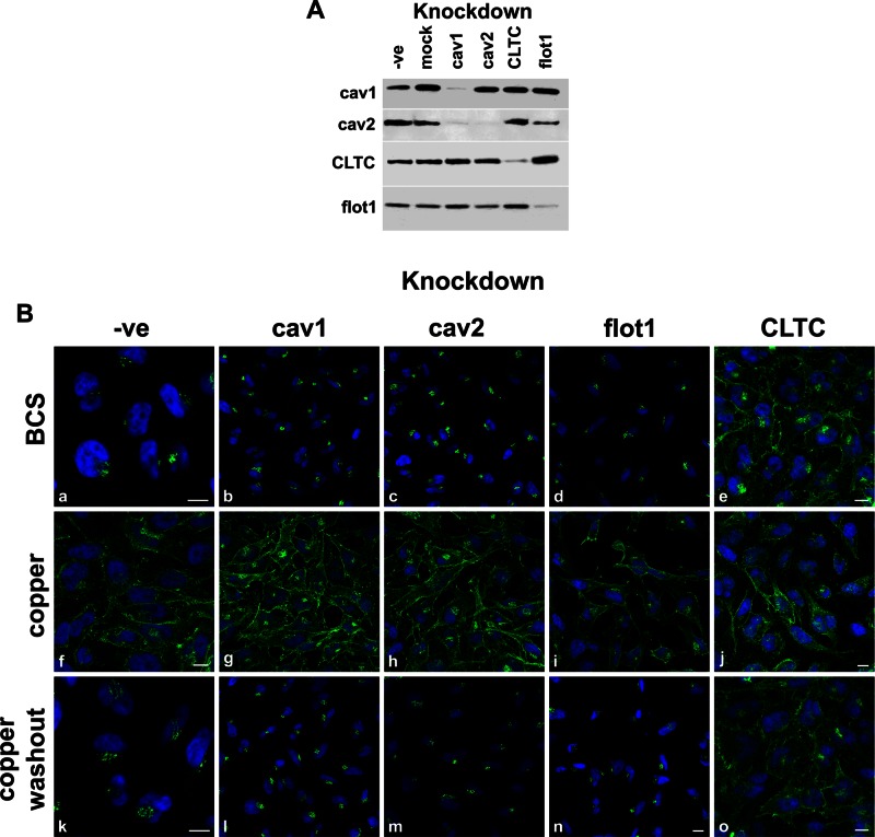
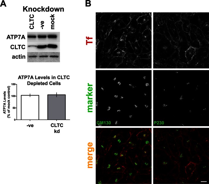
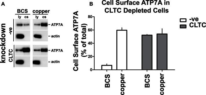
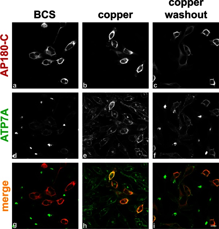
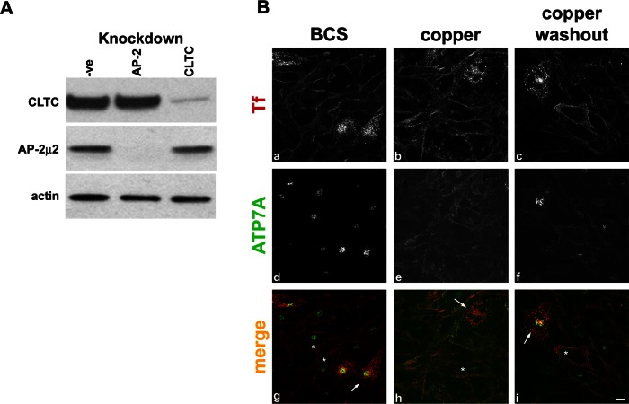
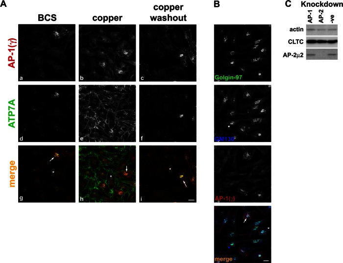
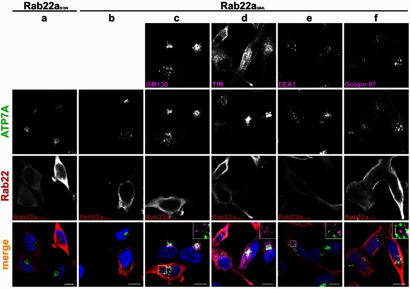
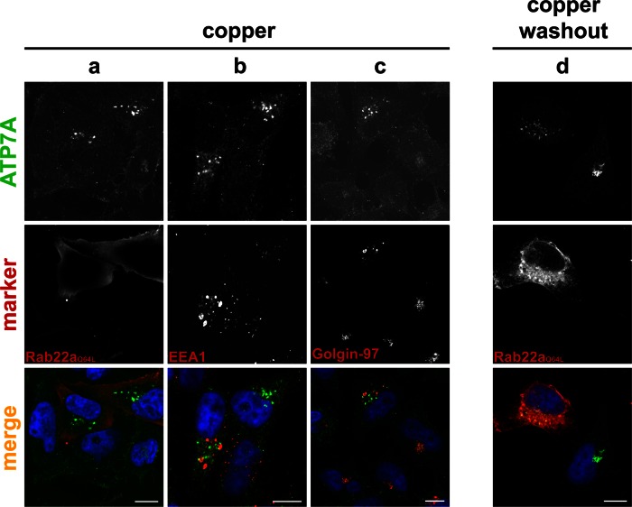
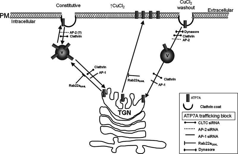
The transporter ATP7A mediates systemic copper absorption and provides cuproenzymes in the trans-Golgi network (TGN) with copper. To regulate metal homeostasis, ATP7A constitutively cycles between the TGN and plasma membrane (PM). ATP7A trafficking to the PM is elevated in response to increased copper load and is reversed when copper concentrations are lowered. Molecular mechanisms underlying this trafficking are poorly understood. We assess the role of clathrin, adaptor complexes, lipid rafts, and Rab22a in an attempt to decipher the regulatory proteins involved in ATP7A cycling. While RNA interference (RNAi)-mediated depletion of caveolin 1/2 or flotillin had no effect on ATP7A localization, clathrin heavy chain depletion or expression of AP180 dominant-negative mutant not only disrupted clathrin-regulated pathways, but also blocked PM-to-TGN internalization of ATP7A. Depletion of the μ subunits of either adaptor protein-2 (AP-2) or AP-1 using RNAi further provides evidence that both clathrin adaptors are important for trafficking of ATP7A from the PM to the TGN. Expression of the GTP-locked Rab22aQ64L mutant caused fragmentation of TGN membrane domains enriched for ATP7A. These appear to be a subdomain of the mammalian TGN, showing only partial overlap with the TGN marker golgin-97. Of importance, ATP7A remained in the Rab22aQ64L-generated structures after copper treatment and washout, suggesting that forward trafficking out of this compartment was blocked. This study provides evidence that multiple membrane-associated factors, including clathrin, AP-2, AP-1, and Rab22, are regulators of ATP7A trafficking.


Figures

FIGURE 1:
FIGURE 1:
Clathrin is required for ATP7A trafficking. HeLa cells were treated with transfection reagent alone (mock), transfected with nonsilencing siRNA (-ve) or siRNA directed against cav1, cav2, flot1, or CLTC. (A) Immunoblot shows depletion of target proteins (indicated above the blot). A total of 10 μg of total protein was loaded per lane. Proteins are detected with the antibodies indicated on the left. (B) Cells were treated with BCS (a–e), CuCl2 (f–j), or CuCl2 followed by washout (k–o) at 37°C before fixation and IF staining with anti-ATP7A antibody (green) and DAPI nuclear stain (blue). Scale bar, 10 μm; where scale bar is not shown, scale is equivalent to that of n.
FIGURE 2:
FIGURE 2:
Clathrin depletion does not affect ATP7A levels or Golgi/TGN architecture. (A) Lysates of siRNA-treated cells containing 10 μg of total protein per lane were separated by SDS–PAGE, followed by immunoblotting with the antibodies indicated on the left of the blot. ATP7A protein levels were quantified, normalized against actin levels, and expressed as percentage of mock control. The graph is representative of six independent experiments. (B) The integrity of the Golgi and the TGN were verified in CLTC-depleted cells by labeling with the cis-Golgi marker GM130 or the TGN marker P230 (green). Cells were incubated with Alexa Fluor 594-Tf (red) at 37°C before washing and fixation to identify clathrin-depleted cells. Scale bar, 10 μm.
FIGURE 3:
FIGURE 3:
Depletion of clathrin causes ATP7A to accumulate at the cell surface. CLTC-deleted or control HeLa cells transfected with nonsilencing siRNA (-ve) were treated with BCS or CuCl2. Cell surface proteins were labeled with biotin at 4°C, followed by cell lysis and isolation of biotinylated cell surface proteins on NeutrAvidin beads. (A) Western blot of prebead lysate (ly) or eluted cell surface e1 protein samples (cs) probed with an antibody to ATP7A or actin. (B) Quantification of cell surface ATP7A, calculated as a percentage of total ATP7A in the cell lysate. The graph is representative of three independent experiments.
FIGURE 4:
FIGURE 4:
ATP7A internalization is inhibited in cells expressing AP180-C. Transiently transected HeLa cells expressing myc-tagged AP180-C were subjected to treatment with BCS, CuCl2, or CuCl2, followed by washout. AP180-C is detected with an antibody against the myc epitope (a–c, g–i red), and cells are counterstained for ATP7A (d–f, g–i green). Scale bar, 10 μm.
FIGURE 5:
FIGURE 5:
Internalization of ATP7A is inhibited in cells depleted of AP-2. (A) Immunoblot comparing lysates from cells treated with nontargeting siRNA (-ve) or depleted of CLTC or AP-2 using siRNA against μ2 subunit of the AP-2 complex. A total of 10 μg of total protein was loaded per lane. Membranes were probed with the antibodies indicated on the left of the blot. (B) HeLa cells depleted of AP-2 were treated with BCS, CuCl2, or CuCl2, followed by washout. Cells showing knockdown of AP-2 are identified by accumulation of Alexa Fluor 594–Tf (a–c, g–i red) at the cell surface, and examples in the merge panels are marked by asterisk. Cells are counterstained for ATP7A (d–f, g–i green). Nondepleted cells showing transferrin internalization are used as an internal negative control; examples are marked with an arrow. Scale bar, 10 μm.
FIGURE 6:
FIGURE 6:
AP-1 depletion blocks ATP7A trafficking to the TGN. (A) HeLa Cells depleted of AP-1 by transfection with siRNA targeting the μ1 subunit were treated with BCS, CuCl2, or CuCl2, followed by washout. Cells depleted of AP-1 are identified by loss of adaptin γ signal at the TGN (a–c, g–i red). Cells are counterstained for ATP7A (d–f, g–i green). Examples of depleted cells are marked in the merge panels with asterisk, and nondepleted cells are marked with an arrow. (B) AP-1–depleted cells, identified by loss of adaptin γ signal (red), are labeled for the trans-Golgi marker golgin‑97 (green) or the cis-Golgi marker GM130 (blue). Scale bar, 10 μm. (C) Immunoblot of lysates from control cells (-ve) or those depleted of AP-1 or AP-2 and blotted with antibodies indicated on the left of the blot. A total of 10 μg of protein was loaded per lane.
FIGURE 7:
FIGURE 7:
Expression of the constitutively active mutant Rab22aQ64L fragments the TGN. HeLa cells transiently transfected with myc-tagged, dominant-negative Rab22aS19N (a) or the constitutively active Rab22aQ64L (b–f) were treated with BCS, fixed, and colabeled with an antibody against myc to detect the Rab22a mutants (a–f red) or with antibodies for the indicated markers (c–f pink). Cells are counterstained for ATP7A (a–f green). DAPI staining (blue) labels the nucleus. ATP7A appears in puncta in Rab22aQ64L-expressing cells. In merge, inset of f, the arrow indicates puncta showing no colocalization between ATP7A and golgin-97 in Rab22aQ64L-expressing cells, and the arrowhead indicates areas of colocalization. Scale bar, 10 μm.
FIGURE 8:
FIGURE 8:
Expression of the Rab22aQ64L mutant inhibits ATP7A trafficking. HeLa cells transiently transfected with myc-tagged, constitutively active Rab22aQ64L were treated with CuCl2, fixed, and colabeled for ATP7A (green) and anti-myc to detect the Rab22a mutant (a), EEA1 (b), or golgin-97 (c) (all red). Cells expressing Rab22aQ64L (d) were treated with copper, followed by washout, and colabeled for ATP7A (green) and myc tag (red). Nuclei are labeled with DAPI (blue). Scale bar, 10 μm.
FIGURE 9:
FIGURE 9:
Regulators of ATP7A trafficking. Schematic representation of the proposed trafficking routes taken by endogenous ATP7A, either constitutively (left) or induced by copper level changes (right). Routes are marked by arrows, and blocks imposed by RNAi, mutant constructs, or drug treatment are shown (see also Table 1). Putative involvement of AP-2 in the constitutive pathway is shown as (?) (see the text). AP180-C shows a similar phenotype to clathrin knockdown and is not included in the scheme. Rab22aQ64L expression causes disruption of the TGN, which is not illustrated. Copper-induced forward pathway is shown as a direct trafficking step to the PM, although intermediary vesicles might be involved as in the constitutive route. V, vesicle.

References

    1. Altschuler Y, Barbas SM, Terlecky LJ, Tang K, Hardy S, Mostov KE, Schmid SL. Redundant and distinct functions for dynamin-1 and dynamin-2 isoforms. J Cell Biol. 1998;143:1871–1881. - PMC - PubMed
    1. Balamurugan K, Schaffner W. Copper homeostasis in eukaryotes: teetering on a tightrope. Biochim Biophys Acta. 2006;1763:737–746. - PubMed
    1. Barral DC, Cavallari M, McCormick PJ, Garg S, Magee AI, Bonifacino JS, De Libero G, Brenner MB. CD1a and MHC class I follow a similar endocytic recycling pathway. Traffic. 2008;9:1446–1457. - PMC - PubMed
    1. Bhatnagar A, Sheffler DJ, Kroeze WK, Compton-Toth B, Roth BL. Caveolin-1 interacts with 5-HT2A serotonin receptors and profoundly modulates the signaling of selected Galphaq-coupled protein receptors. J Biol Chem. 2004;279:34614–34623. - PubMed
    1. Bonifacino JS, Traub LM. Signals for sorting of transmembrane proteins to endosomes and lysosomes. Annu Rev Biochem. 2003;72:395–447. - PubMed

Publication types

MeSH terms