Temporal arteritis: improving patient evaluation with a new protocol
- PMID: 23596371
- PMCID: PMC3627791
- DOI: 10.7812/TPP/12-067
Temporal arteritis: improving patient evaluation with a new protocol
Abstract
Context: The process of diagnosing temporal arteritis remains controversial. Although temporal artery biopsy has long been the standard tool of evaluation, its poor sensitivity has prompted investigation of other methods to aid in diagnosis. Improved clinical evaluation and various imaging techniques have been suggested as ways to establish the diagnosis through noninvasive means and to improve biopsy yield.
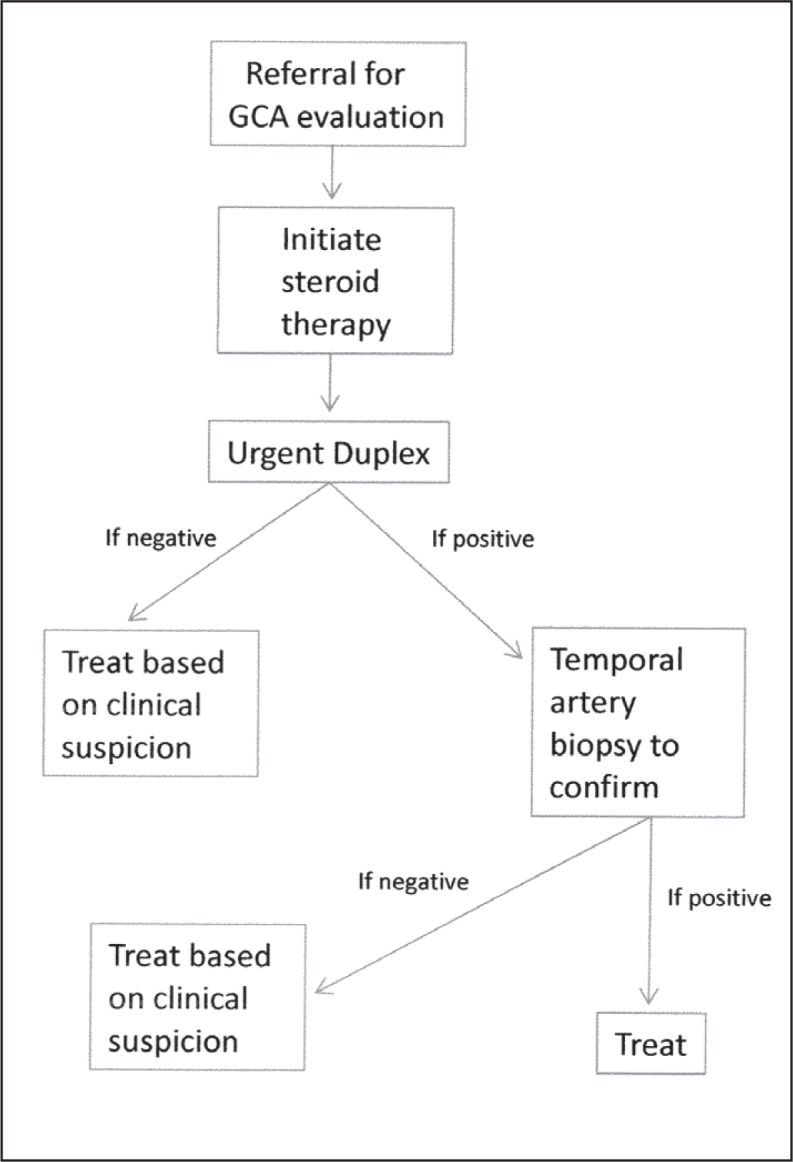
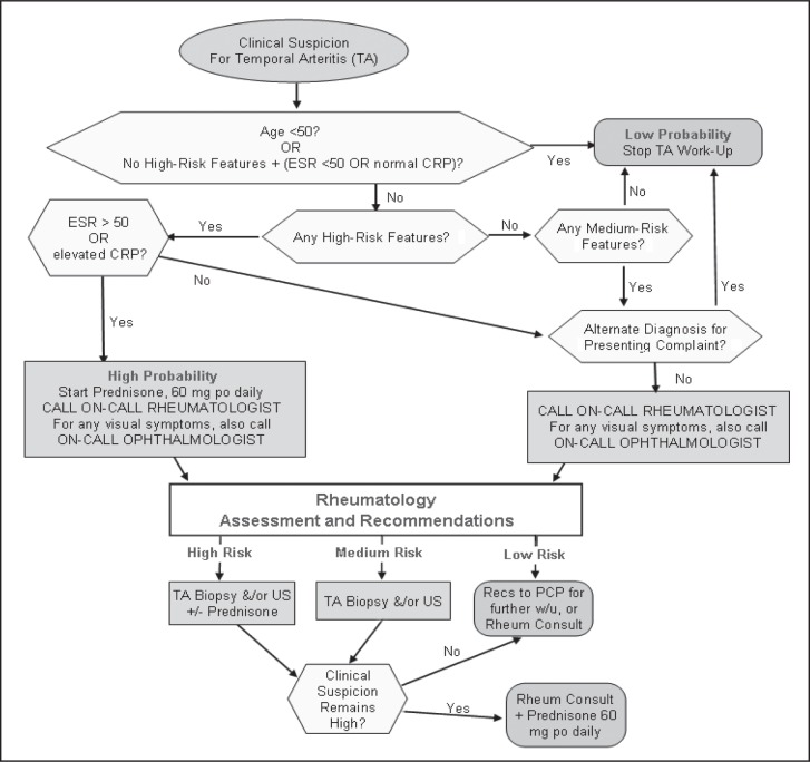
Objective: To retrospectively report and evaluate the process and experience of the Kaiser Permanente Northwest Region in implementing a new protocol that includes an enhanced clinical evaluation as well as the incorporation of color duplex ultrasonography in addition to biopsy when appropriate for temporal arteritis evaluation.
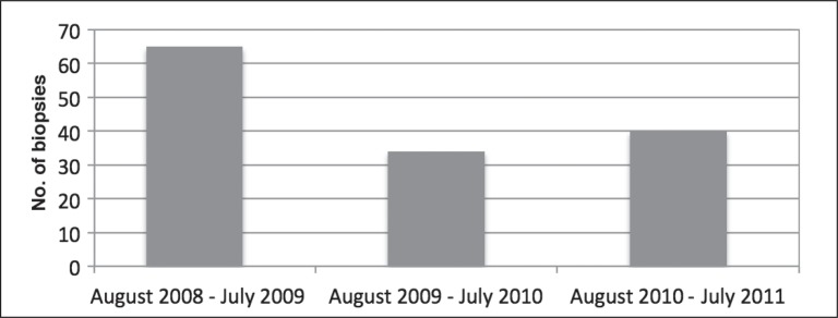
Results: A 38% reduction in the number of temporal artery biopsies performed was achieved through the new protocol, which was created by a multidisciplinary process, including stakeholders from all departments involved. The percentage of abnormal biopsy results rose from 8.5% at baseline to 24%. No cases of the disease were missed after careful evaluation of clinical and medical-legal records.
Conclusions: Adding specialist clinical evaluation and color duplex ultrasonography to the standard diagnostic workup for temporal arteritis creates a rapid, noninvasive, resource-sensible means to diagnose giant cell arteritis, to improve temporal artery biopsy yield, and to decrease the total number of biopsies done. The diagnosis can be made in some cases by clinical evaluation and color duplex ultrasonography alone, thereby saving the patient an unnecessary surgical procedure. Protocols such as this can be implemented by multidisciplinary cooperation in a patient-centered, integrated system.
Figures
References
-
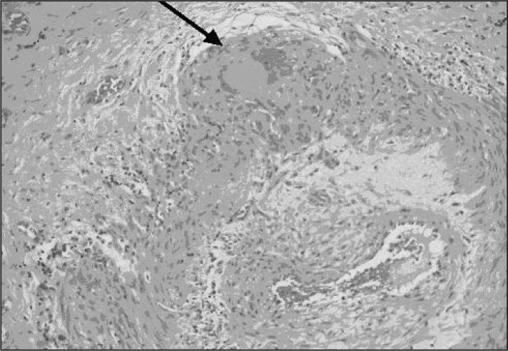
- Mansoor O, Majeed T. A 90 year old woman with painless vision loss. Digital Journal of Ophthalmology. [serial on the Internet] 2005 Feb 10 [cited 2012 Oct 31];11(7): Figure 2. Available from: www.djo.harvard.edu/site.php?url=/physicians/gr/728&page=GR_TT.
-
- González-Gay MA, Garcia-Porrua C, Rivas MJ, Rodriguez-Ledo P, Llorca J. Epidemiology of biopsy proven giant cell arteritis in northwestern Spain: trend over an 18 year period. Ann Rheum Dis. 2001 Apr;60(4):367–71. DOI: http://dx.doi.org/10.1136/ard.60.4.367. - DOI - PMC - PubMed
-
- Butteriss DJ, Clarke L, Dayan M, Birchall D. Use of colour duplex ultrasound to diagnose giant cell arteritis in a case of visual loss of uncertain aetiology. Br J Radiol. 2004 Jul;77(919):607–9. DOI: http://dx.doi.org/10.1259/bjr/22460193. - DOI - PubMed
-
- Schmidt WA, Blockmans D. Use of ultrasonography and positron emission tomography in the diagnosis and assessment of large-vessel vasculitis. Curr Opin Rheumatol. 2005 Jan;17(1):9–15. DOI: http://dx.doi.org/10.1097/01.bor.0000147282.02411.c6. - DOI - PubMed
-
- Narváez J, Narváez JA, Nolla JM, Sirvent E, Reina D, Valverde J. Giant cell arteritis and polymyalgia rheumatica: usefulness of vascular magnetic resonance imaging studies in the diagnosis of aortitis. Rheumatology (Oxford) 2005 Apr;44(4):479–83. DOI: http://dx.doi.org/10.1093/rheumatology/keh513. - DOI - PubMed
MeSH terms
LinkOut - more resources
Full Text Sources
Other Literature Sources
Medical