. 2013 Apr 8:7:34.
doi: 10.3389/fncel.2013.00034. eCollection 2013.

Microglia and monocyte-derived macrophages: functionally distinct populations that act in concert in CNS plasticity and repair

Affiliations

Microglia and monocyte-derived macrophages: functionally distinct populations that act in concert in CNS plasticity and repair

Anat London et al. Front Cell Neurosci. .

Abstract

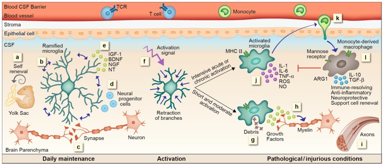
Functional macrophage heterogeneity is recognized outside the central nervous system (CNS), where alternatively activated macrophages can perform immune-resolving functions. Such functional heterogeneity was largely ignored in the CNS, with respect to the resident microglia and the myeloid-derived cells recruited from the blood following injury or disease, previously defined as blood-derived microglia; both were indistinguishably perceived detrimental. Our studies have led us to view the myeloid-derived infiltrating cells as functionally distinct from the resident microglia, and accordingly, to name them monocyte-derived macrophages (mo-MΦ). Although microglia perform various maintenance and protective roles, under certain conditions when they can no longer provide protection, mo-MΦ are recruited to the damaged CNS; there, they act not as microglial replacements but rather assistant cells, providing activities that cannot be timely performed by the resident cells. Here, we focus on the functional heterogeneity of microglia/mo-MΦ, emphasizing that, as opposed to the mo-MΦ, microglia often fail to timely acquire the phenotype essential for CNS repair.

Keywords: CNS; innate; macrophages; microglia; monocyte-derived macrophages; monocytes; neuroprotection; resolution of inflammation.


Figures

FIGURE 1
FIGURE 1
microglial and mo-MΦ functions – cascade of events. (a) Resident microglia originate from yolk sac macrophages that repopulate CNS parenchyma during early development and are self-renewed locally, independent from bone marrow-derived monocytes, by proliferation of primitive progenitors. (b) In the steady state microglia are constantly scanning their environment through their highly motile processes. These cells facilitate the maintenance of synapses (c) and neurogenesis (d), as well as secrete growth factors essential for normal CNS performance (e). Upon recognition of a danger signal, microglia retract their branches, become round and ameboid, and convert into an activated mode (f). A short or moderate signal directs microglia toward a neuroprotective phenotype; these cells clear debris by phagocytosis (g), secrete growth factors associated with remyelination (h) and support regeneration (i). Intensive acute or chronic activation renders microglia neurotoxic; under such conditions microglia fail to acquire a neuroprotective phenotype. Instead, these cells produce reactive oxygen species (ROS), nitric oxide (NO), proteases, and pro-inflammatory cytokines such as IL-1, IL-6, and TNF-α, all of which endanger neuronal activity (j). Microglial malfunction results in the recruitment of mo-MΦ to the damage site (k). mo-MΦ secrete anti-inflammatory cytokines such as IL-10 and TGF-β, express factors associated with immune resolution such as manose receptor and arginase 1 (ARG1), and promote neuroprotection and cell renewal (l), all of which are functions that cannot be provided, under these conditions, by the resident microglia.
FIGURE 2
FIGURE 2
Lessons from other tissue-specific resident macrophages. (A) Langerhans cells (LCs), the resident myeloid cells of the epidermis share with microglia their scanning capacity, their activation mode and possibly, their embryonic origin. Different from microglia these cells migrate to the lymph node where they act as antigen presenting cells. (B) Analogous to microglia, intestinal macrophages act as the first line of defense, protecting the mucosa from harmful pathogens and removing dead cells and debris. Unlike most other tissue macrophages, upon activation by certain stimuli, these cells produce immune-resolving factors. Distinct from microglia, circulating monocytes are largely accepted as the source of intestinal macrophages, however, the possibility of local self-renewal by embryonic precursors, under steady state, was also raised. (C) Kupffer cells are the macrophages of the liver. Similar to microglia, these cells perform clearance of host-related debris and pathogens. Kupffer cells are classical antigen presenting cells; however can also display immune-resolving functions. Moreover, they are largely assumed to be self-renewed independently from circulating monocytes, but a certain Kupffer cell subset was reported to originate from hematogenous precursors.

References

    1. Aarum J., Sandberg K., Haeberlein S. L., Persson M. A. (2003). Migration and differentiation of neural precursor cells can be directed by microglia. Proc. Natl. Acad. Sci. U.S.A. 100 15983–15988 - PMC - PubMed
    1. Ajami B., Bennett J. L., Krieger C., Mcnagny K. M., Rossi F. M. (2011). Infiltrating monocytes trigger EAE progression, but do not contribute to the resident microglia pool. Nat. Neurosci. 14 1142–1149 - PubMed
    1. Ajami B., Bennett J. L., Krieger C., Tetzlaff W., Rossi F. M. (2007). Local self-renewal can sustain CNS microglia maintenance and function throughout adult life. Nat. Neurosci. 10 1538–1543 - PubMed
    1. Arnold L., Henry A., Poron F., Baba-Amer Y., Van Rooijen N. Plonquet, A., et al. (2007). Inflammatory monocytes recruited after skeletal muscle injury switch into antiinflammatory macrophages to support myogenesis. J. Exp. Med. 204 1057–1069 - PMC - PubMed
    1. Auffray C., Sieweke M. H., Geissmann F. (2009). Blood monocytes: development, heterogeneity, and relationship with dendritic cells. Annu. Rev. Immunol. 27 669–692 - PubMed