. 2013 Apr 11:7:6.
doi: 10.3389/fnsys.2013.00006. eCollection 2013.

Transcranial direct-current stimulation increases extracellular dopamine levels in the rat striatum

Affiliations

Transcranial direct-current stimulation increases extracellular dopamine levels in the rat striatum

Tomoko Tanaka et al. Front Syst Neurosci. .

Abstract

Background: Transcranial direct-current stimulation (tDCS) is a non-invasive procedure that achieves polarity-dependent modulation of neuronal membrane potentials. It has recently been used as a functional intervention technique for the treatment of psychiatric and neurological diseases; however, its neuronal mechanisms have not been fully investigated in vivo.

Objective/hypothesis: To investigate whether the application of cathodal or anodal tDCS affects extracellular dopamine and serotonin levels in the rat striatum.

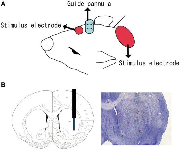
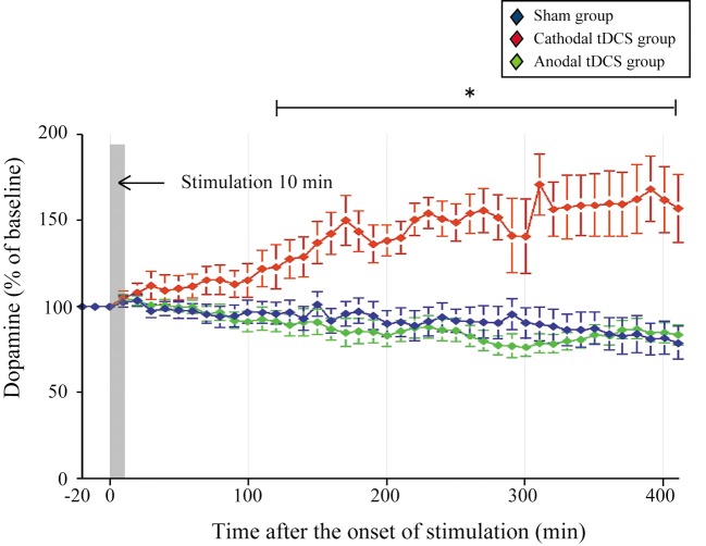
Methods: Stimulation and in vivo microdialysis were carried out under urethane anesthesia, and microdialysis probes were slowly inserted into the striatum. After the collection of baseline fractions in the rat striatum, cathodal or anodal tDCS was applied continuously for 10 min with a current intensity of 800 μA from an electrode placed on the skin of the scalp. Dialysis samples were collected every 10 min until at least 400 min after the onset of stimulation.

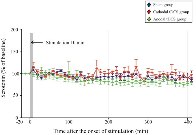
Results: Following the application of cathodal, but not anodal, tDCS for 10 min, extracellular dopamine levels increased for more than 400 min in the striatum. There were no significant changes in extracellular serotonin levels.

Conclusion: These findings suggest that tDCS has a direct and/or indirect effect on the dopaminergic system in the rat basal ganglia.

Keywords: Parkinson disease; basal ganglia; dopamine; striatum; transcranial direct current stimulation.


Figures

Figure 1
Figure 1
Position of tDCS electrode and microdialysis probe. (A) One electrode (small red square) of the stimulator was fixed to the skin with surgical tape just above the brain region including the cortex. The anatomical location corresponded to +2.0 to +7.0 mm anterior and +1.0 to +6.0 mm lateral to the bregma. A second electrode (large red ellipse) was placed on the neck. A guide cannula (blue cylinder) was fixed to the skull with resin dental cement; (B) a microdialysis probe was inserted into the striatum (+1.0 mm anterior, +3.5 mm lateral, and −4.5 mm ventral to the bregma).
Figure 2
Figure 2
Effect of tDCS on extracellular dopamine levels in the striatum. The absolute basal dialysis levels of dopamine in the striatum detected 10 min before the interventions did not differ between groups. Dopamine levels were expressed as percentage signal changes from baseline values before the tDCS application. Group data are presented as the mean ± SEM. Cathodal, but not anodal, tDCS significantly increased extracellular dopamine levels in the striatum. *p < 0.001.
Figure 3
Figure 3
Effect of tDCS on extracellular serotonin levels in the striatum. The absolute basal dialysis levels of serotonin in the striatum detected 10 min before the interventions did not differ between groups. Serotonin levels were expressed as percentage signal changes from baseline values before the tDCS application. Group data are presented as the mean ± SEM. None of the tDCS applications significantly affected extracellular serotonin levels.
Figure 4
Figure 4
Histological examination following tDCS. (A) Cresyl violet staining of the brain tissue below the scalp electrode in the sham group (left) and the cathodal tDCS group (right) 24 h after tDCS application. Scale bars: 1 mm for upper panels, 100 μm for lower panels; (B) HE staining of the skin below the scalp electrode in the sham group (left) and the cathodal tDCS group (right) 24 h after tDCS application. Scale bars: 100 μm for upper panels, 50 μm for lower panels. No morphological change was observed in brain tissue and skin.

Similar articles

Cited by

References

    1. Adcock R. A., Thangavel A., Whitfield-Gabrieli S., Knutson B., Gabrieli J. D. (2006). Reward-motivated learning: mesolimbic activation precedes memory formation. Neuron 50, 507–517 10.1016/j.neuron.2006.03.036 - DOI - PubMed
    1. Alex K. D., Yavanian G. J., McFarlane H. G., Pluto C. P., Pehek E. A. (2005). Modulation of dopamine release by striatal 5-HT2C receptors. Synapse 55, 242–251 10.1002/syn.20109 - DOI - PubMed
    1. Alexander G. E., Crutcher M. D. (1990). Functional architecture of basal ganglia circuits: neural substrates of parallel processing. Trends Neurosci. 13, 266–271 - PubMed
    1. Antal A., Polania R., Schmidt-Samoa C., Dechent P., Paulus W. (2011). Transcranial direct current stimulation over the primary motor cortex during fMRI. Neuroimage 55, 590–596 10.1016/j.neuroimage.2010.11.085 - DOI - PubMed
    1. Bachmann C. G., Muschinsky S., Nitsche M. A., Rolke R., Magerl W., Treede R. D., et al. (2010). Transcranial direct current stimulation of the motor cortex induces distinct changes in thermal and mechanical sensory percepts. Clin. Neurophysiol. 121, 2083–2089 10.1016/j.clinph.2010.05.005 - DOI - PubMed

LinkOut - more resources