. 2013 Apr 15:4:43.
doi: 10.3389/fphar.2013.00043. eCollection 2013.

Connexin mutants and cataracts

Affiliations

Connexin mutants and cataracts

Eric C Beyer et al. Front Pharmacol. .

Abstract

The lens is a multicellular, but avascular tissue that must stay transparent to allow normal transmission of light and focusing of it on the retina. Damage to lens cells and/or proteins can cause cataracts, opacities that disrupt these processes. The normal survival of the lens is facilitated by an extensive network of gap junctions formed predominantly of connexin46 and connexin50. Mutations of the genes that encode these connexins (GJA3 and GJA8) have been identified and linked to inheritance of cataracts in human families and mouse lines. In vitro expression studies of several of these mutants have shown that they exhibit abnormalities that may lead to disease. Many of the mutants reduce or modify intercellular communication due to channel alterations (including loss of function or altered gating) or due to impaired cellular trafficking which reduces the number of gap junction channels within the plasma membrane. However, the abnormalities detected in studies of other mutants suggest that they cause cataracts through other mechanisms including gain of hemichannel function (leading to cell injury and death) and formation of cytoplasmic accumulations (that may act as light scattering particles). These observations and the anticipated results of ongoing studies should elucidate the mechanisms of cataract development due to mutations of lens connexins and abnormalities of other lens proteins. They may also contribute to our understanding of the mechanisms of disease due to connexin mutations in other tissues.

Keywords: cataract; connexin46; connexin50; gap junction; lens.


Figures

Figure 1
Figure 1
Anatomy of the eye (A) and structure of the lens (B). (A) Diagram of the eye illustrating the location of the lens in relation to other structures. The lens is suspended between two transparent fluids, the aqueous and vitreous humors. The aqueous humor fills the space between the cornea and the lens (as well as the posterior chamber). It contains water, oxygen, carbon dioxide, inorganic and organic ions, carbohydrates, glutathione, urea, amino acids, and proteins (including immunoglobulins and growth factors) and is very dynamic (with turnover rates of 1–1.5% per minute) (reviewed by Goel et al., 2010). The aqueous humor stabilizes the ocular structure and contributes to the homeostasis of the avascular structures of the anterior part of the eye by providing nutrition and removing metabolic products. The vitreous is a viscous, gelatinous liquid that fills the space between the lens and the retina. It is essentially a specialized extracellular fluid containing collagen fibers, hyaluronic acid and other soluble proteins, and glycoproteins. The vitreous is rather stagnant, equilibrating very slowly with the plasma. (B) Diagram of the lens showing the distribution of connexin isoforms. Cells from the anterior epithelial cell layer express Cx43 and Cx50, differentiating fiber cells express Cx43, Cx46, and Cx50, and fiber cells (including those of the nucleus) contain Cx46 and Cx50.
Figure 2
Figure 2
Diagrams of hemichannels (A) and intercellular channels (B) formed from a single connexin or two different connexins. (A) Six identical connexin subunits (green or orange) can oligomerize to form a homomeric hemichannel. Two co-expressed connexins may oligomerize with each other to form a heteromeric hemichannel. (B) Two hemichannels dock with each other to form a complete gap junction channel. Two hemichannels of similar composition form homotypic channels whereas two hemichannels of different composition form heterotypic channels.
Figure 3
Figure 3
Diagram illustrating the topology of the human lens connexins, Cx46 (A) and Cx50 (B) and the locations of missense (formula image) and frame shift (formula image) mutations identified in members of families with inherited cataracts. While included in Table 2, Cx50I247M has not been included in the Cx50 diagram, since it may actually be a polymorphism.
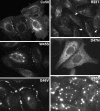
Figure 4
Figure 4
Immunofluorescent localization of wild type Cx50 and of different cataract-associated Cx50 mutants (R23T, W45S, D47N, G46V, and P88S) after their expression by transfection of HeLa cells. Similar to wild type Cx50, W45S and G46V show abundant localization in a punctate distribution along appositional membranes corresponding to gap junction plaques. The abundance of plaques is very reduced for R23T, but small spots at appositional membranes are occasionally observed. D47N and P88S show no localization consistent with gap junction plaque formation. D47N is found in a reticular, cytoplasmic distribution. P88S localizes in intensely fluorescent cytoplasmic inclusions. Reproduced and adapted from Berthoud et al. (2003), Arora et al. (2008), Thomas et al. (2008), and Tong et al. (2011).
Figure 5
Figure 5
Immunofluorescent localization of wild type Cx46, the cataract-associated mutant Cx46fs380 (fs380), Cx46 truncated after amino acid 379 (Tr380) and Cx46fs380 with the FF motif replaced by AA (fs380AA) in transfected HeLa cells. Wild type Cx46 localizes in an intense, linear distribution along appositional membranes as expected for large gap junctions, but such staining is absent for Cx46fs380 which is only found in a cytoplasmic distribution. The cytoplasmic retention must be due to the abnormal sequence in the carboxyl terminus of Cx46fs380, since its removal by truncation (Tr380) restores gap junction formation. Similarly, gap junction formation is restored when the FF motif in Cx46fs380 is replaced with two alanines (fs380AA). Reproduced and adapted from Minogue et al. (2005).
Figure 6
Figure 6
The Cx50G46V mutant induces large hemichannel currents, cytotoxicity, and apoptosis. (A) Hemichannel currents elicited in response to a series of voltage pulses in control Xenopus oocytes (no cRNA) or oocytes injected with cRNA encoding wild type Cx50 or Cx50G46V. The mutant induces much larger currents than wild type Cx50 or control oocytes. (B) Steady-state current-voltage relationships in control (no cRNA), wild type Cx50, and Cx50G46V cRNA-injected oocytes. The mutant induces large outward currents that activate on depolarization. (C–E) Xenopus oocytes were injected with similar amounts of Cx50 (C) or Cx50G46V cRNA (D,E) and incubated in modified Barth’s solution containing 1 mM Ca2+ (C,D) or 3 mM Ca2+ (E) overnight at 18°C. Oocytes injected with Cx50G46V cRNA showed obvious discoloration, membrane disruption, and leakage of yolk (arrows in D) when incubated in modified Barth’s solution containing 1 mM Ca2+ (D) while oocytes injected with Cx50 cRNA showed no apparent detrimental changes when studied under identical conditions (C). The rate of cell death of the Cx50G46V cRNA-injected oocytes was significantly reduced by increasing the external calcium concentration from 1 to 3 mM (E). (F,G). Graphs show the results of cell cycle analysis of propidium iodide-stained HeLa cells induced to express wild type Cx50 (F) or Cx50G46V (G). This analysis revealed a substantial population of cells in the sub-G1 fraction, a marker for apoptosis, in cells expressing Cx50G46V but not in cells expressing Cx50 implying that expression of this mutant increased the proportion of apoptotic cells. Reproduced and adapted from Minogue et al. (2009).

Similar articles

Cited by

References

    1. Abrams C. K., Scherer S. S. (2012). Gap junctions in inherited human disorders of the central nervous system. Biochim. Biophys. Acta 1818, 2030–204710.1016/j.bbamem.2011.08.015 - DOI - PMC - PubMed
    1. Addison P. K., Berry V., Holden K. R., Espinal D., Rivera B., Su H., et al. (2006). A novel mutation in the connexin 46 gene (GJA3) causes autosomal dominant zonular pulverulent cataract in a Hispanic family. Mol. Vis. 12, 791–795 - PubMed
    1. Alapure B. V., Stull J. K., Firtina Z., Duncan M. K. (2012). The unfolded protein response is activated in connexin 50 mutant mouse lenses. Exp. Eye Res. 102, 28–3710.1016/j.exer.2012.06.004 - DOI - PMC - PubMed
    1. Arora A., Minogue P. J., Liu X., Addison P. K., Russel-Eggitt I., Webster A. R., et al. (2008). A novel connexin50 mutation associated with congenital nuclear pulverulent cataracts. J. Med. Genet. 45, 155–16010.1136/jmg.2007.051029 - DOI - PMC - PubMed
    1. Arora A., Minogue P. J., Liu X., Reddy M. A., Ainsworth J. R., Bhattacharya S. S., et al. (2006). A novel GJA8 mutation is associated with autosomal dominant lamellar pulverulent cataract: further evidence for gap junction dysfunction in human cataract. J. Med. Genet. 43, e2.10.1136/jmg.2005.034108 - DOI - PMC - PubMed

LinkOut - more resources