. 2013 Apr 15;8(4):e61407.
doi: 10.1371/journal.pone.0061407. Print 2013.

The C-type lectin of the aggrecan G3 domain activates complement

Affiliations

The C-type lectin of the aggrecan G3 domain activates complement

Camilla Melin Fürst et al. PLoS One. .

Abstract

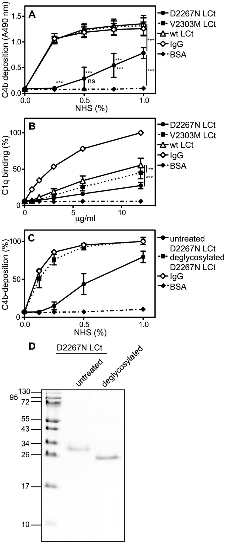
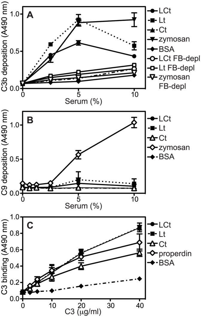
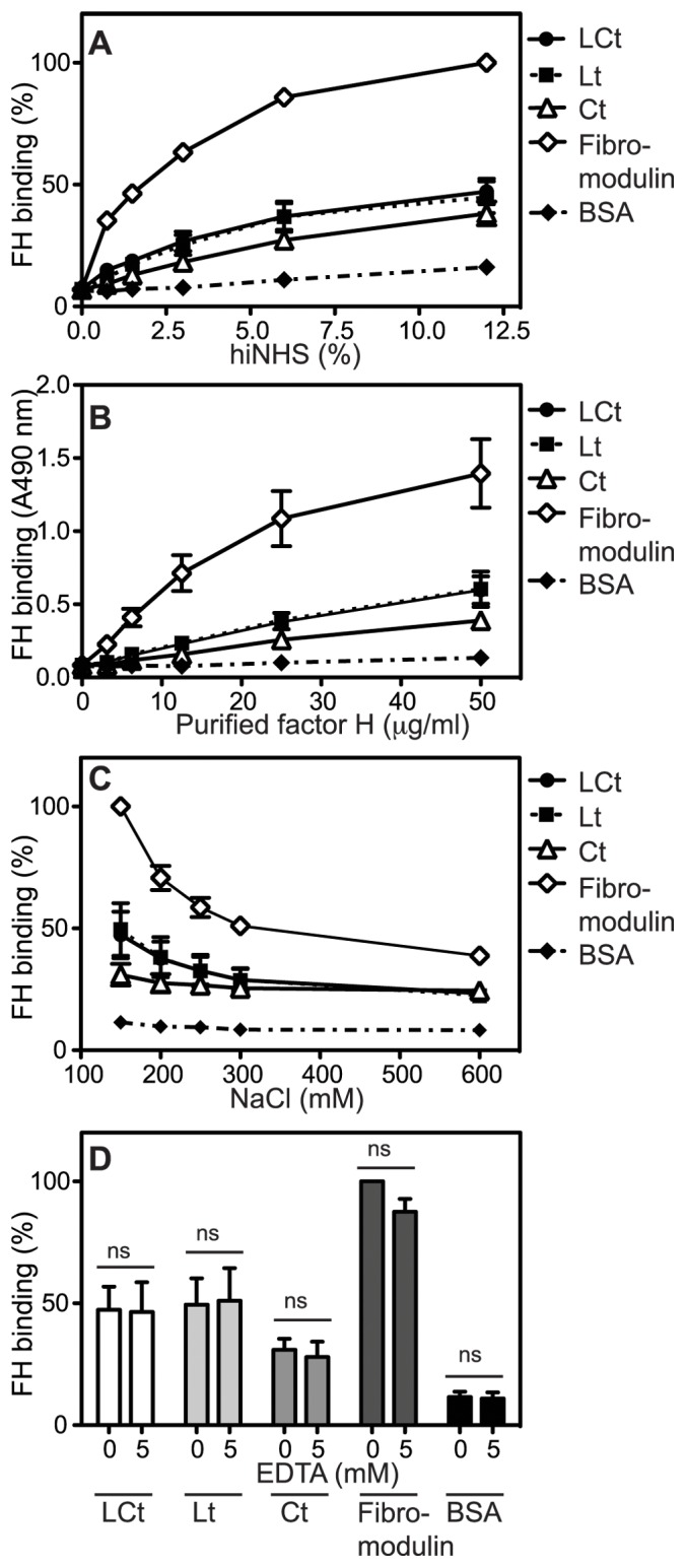
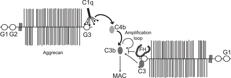
Excessive complement activation contributes to joint diseases such as rheumatoid arthritis and osteoarthritis during which cartilage proteins are fragmented and released into the synovial fluid. Some of these proteins and fragments activate complement, which may sustain inflammation. The G3 domain of large cartilage proteoglycan aggrecan interacts with other extracellular matrix proteins, fibulins and tenascins, via its C-type lectin domain (CLD) and has important functions in matrix organization. Fragments containing G3 domain are released during normal aggrecan turnover, but increasingly so in disease. We now show that the aggrecan CLD part of the G3 domain activates the classical and to a lesser extent the alternative pathway of complement, via binding of C1q and C3, respectively. The complement control protein (CCP) domain adjacent to the CLD showed no effect on complement initiation. The binding of C1q to G3 depended on ionic interactions and was decreased in D2267N mutant G3. However, the observed complement activation was attenuated due to binding of complement inhibitor factor H to CLD and CCP domains. This was most apparent at the level of deposition of terminal complement components. Taken together our observations indicate aggrecan CLD as one factor involved in the sustained inflammation of the joint.


Conflict of interest statement

Competing Interests: The authors have declared that no competing interests exist.

Figures

Figure 1
Figure 1. Structure of aggrecan and localization of G3 domain fragments which were used in the study.
A, schematic outline of human aggrecan, including three globular domains (G1–G3), which is involved in numerous interactions with other ECM components. The carboxyl-terminal globular G3 domain consists of one or two EGF-like domains, a C-type lectin domain (CLD) and a complement control protein (CCP) domain followed by a short tail. In the current study we used three wild-type recombinant fragments of G3 domain: LCt, Lt and Ct and two mutants of LCt containing D2267N or V2303M substitutions. B, the wild-type and mutated fragments of G3 domain were expressed in eukaryotic cells and separated by SDS/PAGE followed by Coomassie Blue staining.
Figure 2
Figure 2. Lectin domain of aggrecan G3 module activates the classical pathway.
Aggregated IgG (positive control for classical pathway), LCt, Lt, Ct and BSA (negative control) were coated onto microtiter plates and increasing concentrations of normal human serum (NHS) were added. Activation of the classical pathway was measured by detecting deposited C1q (A), C4b (B), C3b (C) and C9 (D). The graphs show the mean and standard deviation (SD) of three independent experiments. Statistical significance of differences was calculated using a two-way ANOVA with a Bonferroni posttest. * p<0.05, **p<0.01, ***p<0.001.).
Figure 3
Figure 3. Lectin domain of aggrecan G3 domain activates classical pathway due to binding of C1q.
A, aggregated IgG (positive control for classical pathway), mannan (positive control for lectin pathway), LCt, Lt, Ct and BSA (negative control) were coated onto microtiter plates and increasing concentrations of C1q-depleted human serum were added. Activation of the classical pathway was measured by detecting deposited C4b. The absorbance was normalized relative to deposition on mannan from 10% C1q-depleted serum. B, aggregated IgG, LCt, Lt, Ct and BSA were coated onto microtiter plates and incubated with purified C1q, which was then detected with specific antibodies. Aggregated IgG, LCt, Lt, Ct and BSA were coated onto microtiter plates and incubated with purified C1q in a buffer supplemented with increasing NaCl concentrations (C) or EDTA (D). The graphs show the mean and SD of three independent experiments. Statistical significance of differences was calculated using two-way ANOVA with Bonferroni posttest (B, D). (*p<0.05, **p<0.01, ***p<0.001.).
Figure 4
Figure 4. C1q binds the G3 domain and a part of the aggrecan core protein.
A–E, using electron microscopy, complexes between bovine nasal cartilage aggrecan and C1q were visualized by negative staining. G1 and G3 domains of aggrecan were labelled with antibody-conjugated colloidal gold (10 nm and 5 nm, respectively) and are pointed out by black (G1) and white (G3) arrowheads. Scale bar shows 100 nm (A) and 50 nm (B). A, several aggrecan molecules bound to the heads of C1q through their G3 domains. C1q also bound the core of aggrecan. B–D, selected complexes between single molecules of C1q and one to three aggrecan molecules through their G3 domains, at a higher magnification. E, single molecule of C1q bound to the core of aggrecan. F, microtiter plates were coated with aggregated IgG (positive control), adult, calf and fetal bovine articular cartilage aggrecan and BSA (negative control) and deposited C4b from increasing concentrations of NHS was detected. The graph show the mean and SD of three independent experiments. Statistical significance of differences was calculated using two-way ANOVA with Bonferroni posttest. (*p<0.05, **p<0.01, ***p<0.001.).
Figure 5
Figure 5. The mutation D2267N within the G3 domain decreases the complement activation capacity.
A–B, microtiter plates were coated with aggregated IgG (positive control for classical pathway), LCt carrying the mutation D2267N or V2303M, wt LCt, and BSA (negative control). A, increasing concentrations of NHS were added and complement activation was measured by detecting deposited C4b. B, plates were incubated with purified C1q, which was then detected with specific antibodies. C, Microtiter plates were coated with untreated or N-glycosidase F-treated D2267N LCt, aggregated IgG and BSA. Proteins were incubated with increasing concentrations of NHS and deposited C4b was detected. Note that in addition to deglycosylation, the deamidation resulting from N-glycosidase F treatment reverts the protein sequence to wild type. D, D2267N LCt was deglycosylated with N-glycosidase F and untreated and deglycosylated proteins were separated by SDS/PAGE followed by Coomassie Blue staining. The graphs show the mean and SD of three independent experiments. Statistical significance of differences was calculated using two-way ANOVA with Bonferroni posttest. (*p<0.05, **p<0.01, ***p<0.001.).
Figure 6
Figure 6. Lectin domain of aggrecan G3 module triggers limited activation of the alternative pathway.
Zymosan (positive control for alternative pathway), LCt, Lt, Ct and BSA (negative control) were coated onto microtiter plates and increasing concentrations of NHS or factor B-depleted (FB-depl) human serum in Mg++EGTA were added. Activation of the alternative pathway was measured by detecting deposited C3b (A) and C9 (B). Properdin (positive control for binding of C3), LCt, Lt, Ct and BSA (negative control) were coated onto microtiter plates and incubated with purified C3, which was then detected with specific antibodies (C). The graphs show the mean and SD of three independent experiments.
Figure 7
Figure 7. Lectin domain of aggrecan G3 module binds factor H (FH).
Microtiter plates were coated with fibromodulin (positive control for binding of FH), LCt, Lt, Ct and BSA (negative control) and the binding of FH from heat-inactivated NHS (hi-NHS) (A) or solution containing purified FH (B) was detected using specific antibodies. Fibromodulin, LCt, Lt, Ct and BSA were coated onto microtiter plates and incubated with purified FH in a buffer supplemented with increasing NaCl concentrations (C) or EDTA (D). In A, C and D absorbance was normalized relative to the highest absorbance value obtained with fibromodulin in each figure. The graphs show the mean and SD of three independent experiments. The differences in binding using different EDTA concentrations (D) were not statistically significant, calculated using two-way ANOVA with Bonferroni posttest. (*, p<0.05, **, p<0.01, ***, p<0.001.).
Figure 8
Figure 8. Schematic representation of interactions between aggrecan G3 domain and complement.
C1-complex binds the aggrecan G3 domain via the globular heads of C1q, which leads to activation of the classical pathway and the deposition of C4b. Classical pathway C3-convertase is formed, which generates C3b, which is further enhanced by the alternative pathway acting as amplification loop. Furthermore, interaction of G3 with C3 leads to low degree activation of the alternative pathway. Deposited C3b is included in C3- and C5 convertases that finally lead to the formation of MAC. Complement factor FH also binds G3 and is suggested to attenuate the amount of deposited C3b and formed MAC.

Similar articles

Cited by

References

    1. Ricklin D, Hajishengallis G, Yang K, Lambris JD (2010) Complement: a key system for immune surveillance and homeostasis. Nat Immunol 11: 785–797. - PMC - PubMed
    1. Sjoberg AP, Trouw LA, Blom AM (2009) Complement activation and inhibition: a delicate balance. Trends Immunol 30: 83–90. - PubMed
    1. Okroj M, Heinegard D, Holmdahl R, Blom AM (2007) Rheumatoid arthritis and the complement system. Ann Med 39: 517–530. - PubMed
    1. Kojouharova M, Reid K, Gadjeva M (2010) New insights into the molecular mechanisms of classical complement activation. Mol Immunol 47: 2154–2160. - PubMed
    1. Groeneveld TW, Oroszlan M, Owens RT, Faber-Krol MC, Bakker AC, et al. (2005) Interactions of the extracellular matrix proteoglycans decorin and biglycan with C1q and collectins. J Immunol 175: 4715–4723. - PubMed

Publication types

MeSH terms

LinkOut - more resources