

Review
doi: 10.1016/j.rdc.2013.02.007.
Epub 2013 Mar 13.
Extrapulmonary manifestations of sarcoidosis
Affiliations
- PMID: 23597964
- PMCID: PMC3756667
- DOI: 10.1016/j.rdc.2013.02.007
Item in Clipboard
Review
Extrapulmonary manifestations of sarcoidosis
Rheum Dis Clin North Am.
2013 May.
Abstract
Sarcoidosis is a systemic disease characterized by the development of epithelioid granulomas in various organs. Although the lungs are involved in most patients with sarcoidosis, virtually any organ can be affected. Recognition of extrapulmonary sarcoidosis requires awareness of the organs most commonly affected, such as the skin and the eyes, and vigilance for the most dangerous manifestations, such as cardiac and neurologic involvement. In this article, the common extrapulmonary manifestations of sarcoidosis are reviewed and organ-specific therapeutic considerations are discussed.
Copyright © 2013 Elsevier Inc. All rights reserved.
Figures

Examples of cutaneous sarcoidosis. (A) Waxy papules over the eyelid of a patient with systemic sarcoidosis. (B) Granulomatous inflammation within the area of 1 color of a tattoo in a patient with systemic sarcoidosis. (Courtesy of Dr J. Merola, Brigham and Women’s Hospital, Boston, MA.)

Examples of ocular sarcoidosis. (A) Scleritis in a patient with sarcoidosis. (B) Optic nerve swelling on fundoscopic exam. (Courtesy of Dr G. Papaliodis, Massachusetts Eye and Ear Infirmary, Boston, MA.)
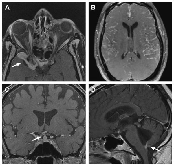
Examples of neurosarcoidosis. (A) Orbital and dural involvement (arrow) on gadolinium-enhanced T1-weighted fat-saturated MRI. (B) Diffuse nodular meningeal lesions on gadolinium-enhanced T1-weighted fat-saturated MRI. (C) Involvement of the pituitary infundibulum (arrow) on gadolinium-enhanced T1-weighted fat-saturated MRI. (D) Involvement of the foramen of Magendie (arrow) causing hydrocephalus. (Courtesy of Dr K. Talekar, Thomas Jefferson University Hospital, Philadelphia, PA.)
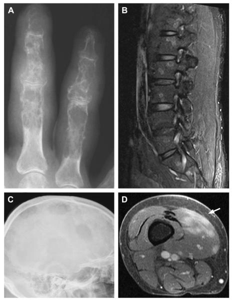
Examples of musculoskeletal involvement in sarcoidosis. (A) Punched-out cortical lesions and coarsened trabeculae yield a characteristic lacelike pattern in the proximal, middle, and distal phalanges. (B) Focal targetlike lesions in the vertebral bodies seen on sagittal short-tau inversion recovery sequence MRI. (C) Multiple large, lytic lesions within the skull of a patient with sarcoidosis. (D) Lobular lesions (arrow) within the vastus medialis muscle on gadolinium-enhanced MRI. (Courtesy of [A, C, D] Dr S. Smith, Brigham and Women’s Hospital, Boston, MA; and [B] Dr K. Talekar, Thomas Jefferson University Hospital, Philadelphia, PA.)
References
-
- Statement on sarcoidosis. Joint Statement of the American Thoracic Society (ATS), the European Respiratory Society (ERS) and the World Association of Sarcoidosis and Other Granulomatous Disorders (WASOG) adopted by the ATS Board of Directors and by the ERS Executive Committee, February 1999. Am J Respir Crit Care Med. 1999;160:736–55. - PubMed
-
- Iannuzzi MC, Rybicki BA, Teirstein AS. Sarcoidosis. N Engl J Med. 2007;357:2153–65. - PubMed
-
- Pietinalho A, Hiraga Y, Hosoda Y, et al. The frequency of sarcoidosis in Finland and Hokkaido, Japan. A comparative epidemiological study. Sarcoidosis. 1995;12:61–7. - PubMed
-
- Rybicki BA, Major M, Popovich J, Jr, et al. Racial differences in sarcoidosis incidence: a 5-year study in a health maintenance organization. Am J Epidemiol. 1997;145:234–41. - PubMed
Publication types
MeSH terms
Substances
Grants and funding
LinkOut - more resources
Full Text Sources
Other Literature Sources
Medical
