. 2014 Apr;259(4):715-22.
doi: 10.1097/SLA.0b013e31828fae14.

Intestinal alkaline phosphatase prevents antibiotic-induced susceptibility to enteric pathogens

Affiliations

Intestinal alkaline phosphatase prevents antibiotic-induced susceptibility to enteric pathogens

Sayeda Nasrin Alam et al. Ann Surg. 2014 Apr.

Abstract

Objective: To determine the efficacy of oral supplementation of the gut enzyme intestinal alkaline phosphatase (IAP) in preventing antibiotic-associated infections from Salmonella enterica serovar Typhimurium (S. Typhimurium) and Clostridium difficile.

Background: The intestinal microbiota plays a pivotal role in human health and well-being. Antibiotics inherently cause dysbiosis, an imbalance in the number and composition of intestinal commensal bacteria, which leads to susceptibility to opportunistic bacterial infections. Previously, we have shown that IAP preserves the normal homeostasis of intestinal microbiota and that oral supplementation with calf IAP (cIAP) rapidly restores the normal gut flora. We hypothesized that oral IAP supplementation would protect against antibiotic-associated bacterial infections.

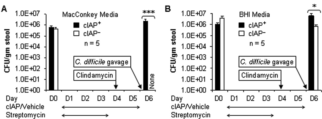
Methods: C57BL/6 mice were treated with antibiotic(s) ± cIAP in the drinking water, followed by oral gavage of S. Typhimurium or C. difficile. Mice were observed for clinical conditions and mortality. After a defined period of time, mice were killed and investigated for hematological, inflammatory, and histological changes.

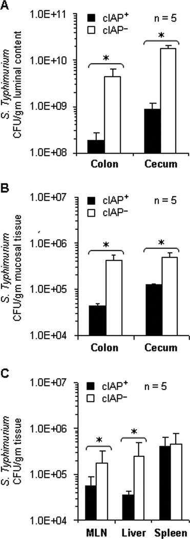
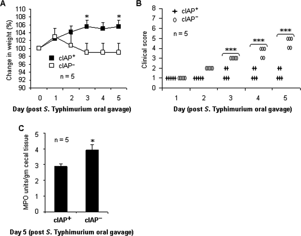
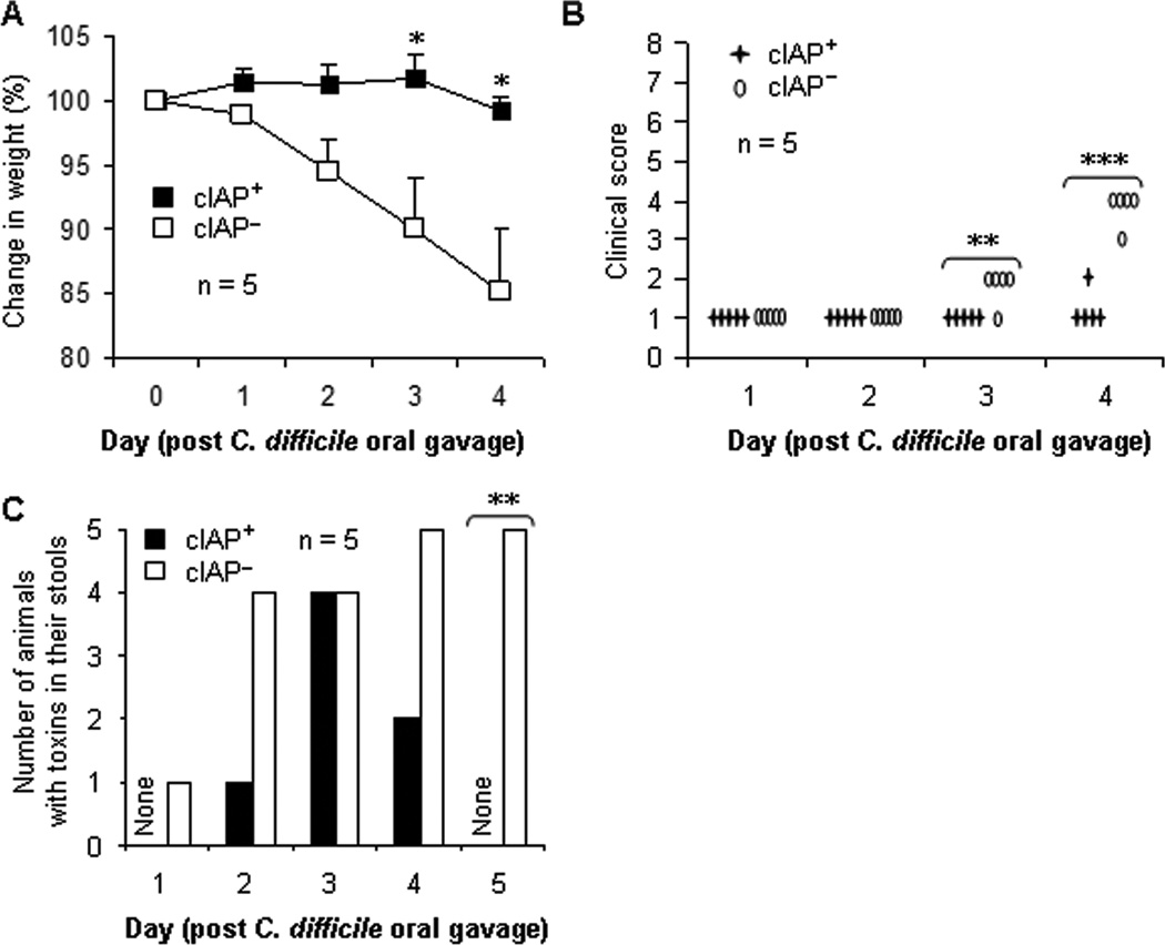
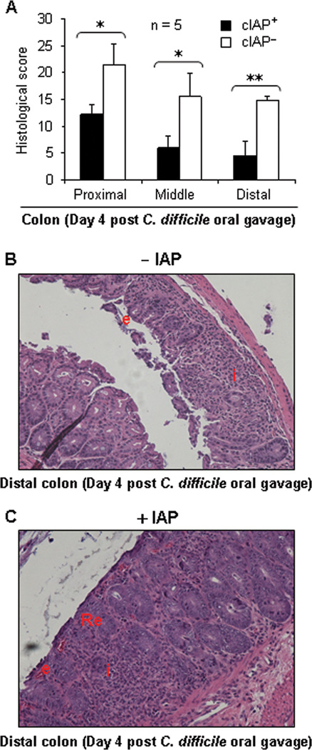
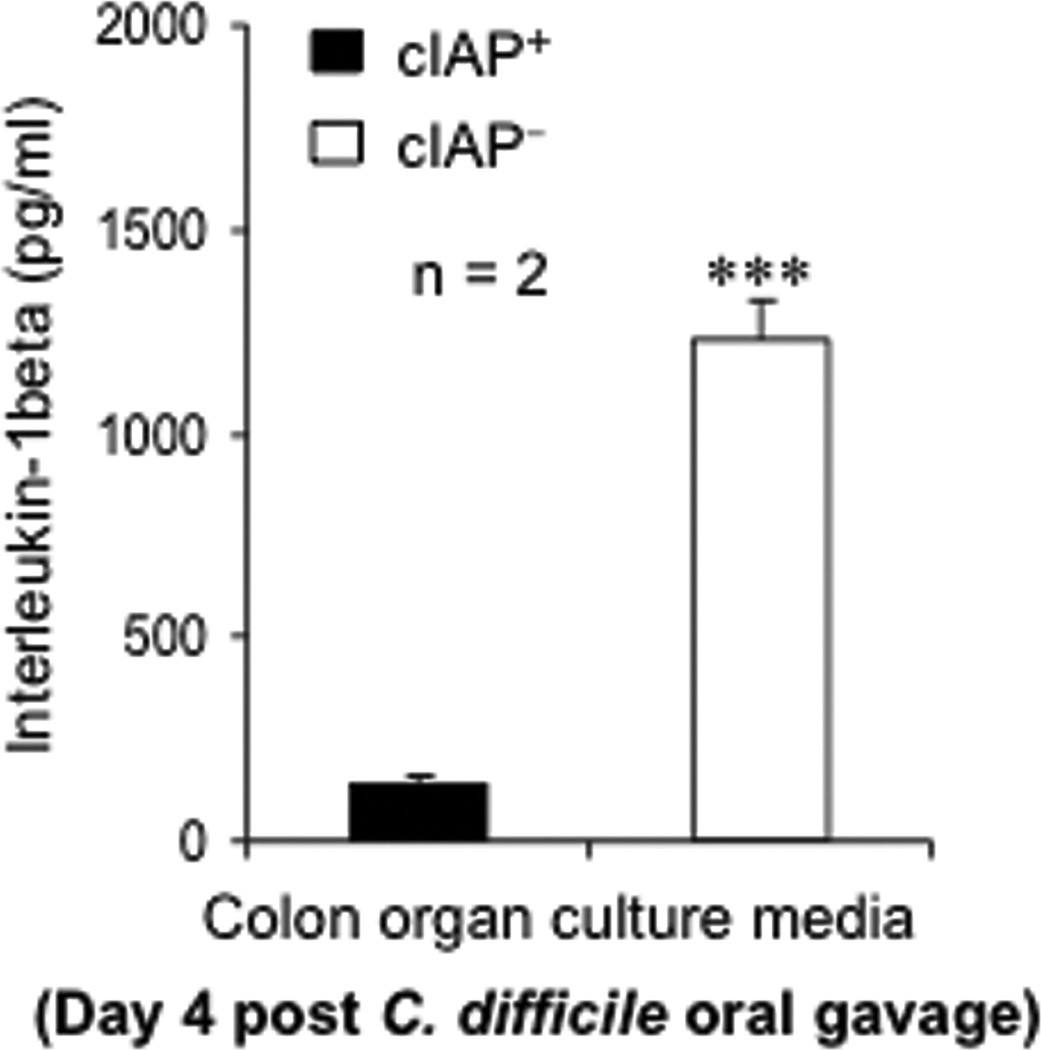
Results: We observed that oral supplementation with cIAP during antibiotic treatment protects mice from infections with S. Typhimurium as well as with C. difficile. Animals given IAP maintained their weight, had reduced clinical severity and gut inflammation, and showed improved survival.

Conclusions: Oral IAP supplementation protected mice from antibiotic-associated bacterial infections. We postulate that oral IAP supplementation could represent a novel therapy to protect against antibiotic-associated diarrhea (AAD), C. difficile-associated disease (CDAD), and other enteric infections in humans.


Conflict of interest statement

Competing interests: The authors declare no conflicting financial interests.

Figures

Figure 1
Figure 1
Oral Supplementation with cIAP Reduces Intestinal Colonization and Systemic Translocation of Salmonella Typhimurium. Groups of mice (n = 5) were co-treated with streptomycin (5 mg/ml) +/− cIAP (200 U/ml) in the drinking water for 3 days. Two days after discontinuation of streptomycin, mice were gavaged with 5,000 colony forming units (CFU) of S. Typhimurium. cIAP treatment continued for another 5 days (total 10 days). Mice were sacrificed 5 days after bacterial gavage and CFU in different tissues were determined. Values are expressed as mean +/− SEM. *, p < 0.05 (Student’s t test). (A) S. Typhimurium CFU in the luminal content of colon and cecum. (B) S. Typhimurium CFU in the mucosa of colon and cecum. (C) S. Typhimurium CFU in the mesenteric lymph nodes (MLN), liver and spleen.
Figure 2
Figure 2
Oral IAP Supplementation Improves Clinical Conditions in Mice Infected with S. Typhimurium. For the description of animals see Figure 1 and also Methods. Mice were observed for weight and clinical conditions for 5 days after bacterial gavage. For determining myeloperoxidase (MPO) activity cecal tissue was homogenized by sonication and supernatant was collected after centrifugation followed by MPO assay. MPO activity was expressed as average units/gm cecal tissue +/− SD. (A) Percentage change in weight of mice infected with S. Typhimurium and treated with cIAP (cIAP+) or without cIAP (cIAP). *, p < 0.05 (Student’s t test). (B) Clinical score of mice infected with S. Typhimurium and treated with cIAP (cIAP+) or without cIAP (cIAP). ***, p < 0.001 (Student’s t test). (C) MPO activity in the cecal tissue of mice infected with S. Typhimurium and treated with cIAP (cIAP+) or without cIAP (cIAP). *, p < 0.05; ***, p < 0.001 (Student’s t test).
Figure 3
Figure 3
Oral Supplementation with cIAP Rapidly Restores the Normal Gut Flora in Mice after Discontinuation of Treatment with Multiple Antibiotics. Groups of mice were co-treated with antibiotics (streptomycin 3 days orally followed by a single intraperitoneal injection of clindamycin on day 4) +/− cIAP (200 U/ml). Individual stool samples were grown on MacConkey agar plates (A) and Brain Heart Infusion (BHI) agar plates (B). Please note that on days 2 to 5 animals from both groups did not grow any bacterial colonies from the collected stool samples indicating the effectiveness of antibiotic treatment. Values are expressed as average CFU/gm stool +/− SEM. *, p < 0.05; ***, p < 0.001 (Student’s t test).
Figure 4
Figure 4
Oral cIAP Supplementation Improves Clinical Conditions in Mice Infected with C. difficile. For the description of animals see Figure 3 and also Methods. Groups of mice were co-treated with antibiotics (streptomycin 3 days orally followed by a single intraperitoneal injection of clindamycin on day 4) +/− cIAP (200 U/ml). On day 6, IAP/vehicle was discontinued and both groups received approximately 1,000 CFU of C. difficile by oral gavage. Weight, clinical conditions and the presence of C. difficile toxins in the stool were monitored daily. (A) Percentage change in weight of mice infected with C. difficile and treated with cIAP (cIAP+) or without cIAP (cIAP). *, p < 0.05 (Student’s t test). (B) Clinical score of mice infected with C. difficile and treated with cIAP (cIAP+) or without cIAP (cIAP). **, p < 0.01; ***, p < 0.001 (Student’s t test). (C) Clearance of toxins in mice infected with C. difficile and treated with cIAP (cIAP+) or without cIAP (cIAP). **, p < 0.01 (Student’s t test).
Figure 5
Figure 5
Oral Supplementation with cIAP Minimizes Colonic Injury in C. difficile-Infected Mice. For the description of animals see Figure 3 and also Methods. Groups of mice were co-treated with antibiotics (streptomycin 3 days orally followed by a single intraperitoneal injection of clindamycin on day 4) +/− cIAP (200 U/ml). On day 6, IAP/vehicle was discontinued and both groups received approximately 1,000 CFU of C. difficile by oral gavage. Mice were sacrificed 4 days after C. difficile infection. Hematoxylin and Eosin (H&E) staining was performed on slides from different segments of the colon and the mucosa examined. Lesions were graded based on an established grading system. (A) Histological score of mice infected with C. difficile and treated with cIAP (cIAP+) or without cIAP (cIAP). Note: The lower the score the better is the outcome. *, p < 0.05; **, p < 0.01 (Student’s t test). (B) Representative histological slide of distal colon of C. difficile-infected mice not treated with cIAP (−IAP). (C) Representative histological slide of distal colon of C. difficile-infected mice treated with cIAP (+IAP). Structures: e, epithelial disruption; i, inflammatory infiltrate; Re, re-epithelialization. Magnification: 200×. Note: Extensive epithelial disruption (e) is evident in the untreated mice (−IAP).
Figure 6
Figure 6
Oral Supplementation with cIAP Minimizes Inflammation. Mice were treated with cIAP (cIAP+) or without cIAP (cIAP) for 5 days and then infected with C. difficile. For the description of animals see Figure 3 and also Methods. Animals were sacrificed 4 days after C. difficile infection. Colonic segments were obtained and organ culture performed. Using an ELISA kit, proinflammatory cytokine interleukin-1beta (IL-1β) levels were determined in the colon organ culture media. Values are expressed as pg/ml culture media +/− SD. ***, p < 0.001; (Student’s t test).

Similar articles

Cited by

References

    1. Miller CP, Bohnhoff M. Changes in the Mouse's Enteric Microflora Associated with Enhanced Susceptibility to Salmonella Infection Following Streptomycin Treatment. J Infect Dis. 1963;113:59–66. - PubMed
    1. van der Waaij D, Berghuis-de Vries JM, Lekkerkerk L-v. Colonization resistance of the digestive tract in conventional and antibiotic-treated mice. J Hyg (Lond) 1971;69(3):405–411. - PMC - PubMed
    1. Sekirov I, Tam NM, Jogova M, et al. Antibiotic-induced perturbations of the intestinal microbiota alter host susceptibility to enteric infection. Infect Immun. 2008;76(10):4726–4736. - PMC - PubMed
    1. Chen X, Katchar K, Goldsmith JD, et al. A mouse model of Clostridium difficile-associated disease. Gastroenterology. 2008;135(6):1984–1992. - PubMed
    1. Glynn MK, Reddy V, Hutwagner L, et al. Prior antimicrobial agent use increases the risk of sporadic infections with multidrug-resistant Salmonella enterica serotype Typhimurium: a FoodNet case-control study, 1996–1997. Clin Infect Dis. 2004;38(Suppl 3):S227–S236. - PubMed

Publication types

MeSH terms