Review
. 2014 Apr;35(4):625-31.
doi: 10.3174/ajnr.A3489. Epub 2013 Apr 18.

The role of MR imaging in assessment of brain damage from carbon monoxide poisoning: a review of the literature

Affiliations
Review

The role of MR imaging in assessment of brain damage from carbon monoxide poisoning: a review of the literature

T Beppu. AJNR Am J Neuroradiol. 2014 Apr.

Abstract

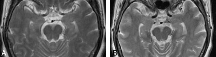
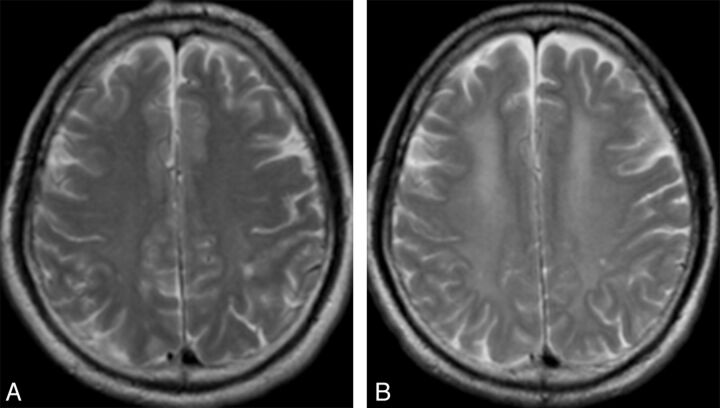
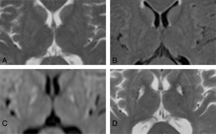
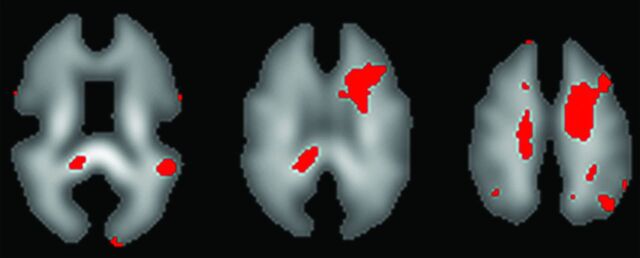
The aim of this article is to review how MR imaging and associated imaging modalities provide clinicopathologic information on brain damage after carbon monoxide poisoning. Initially, many authors documented typical findings of conventional MR imaging in the gray matter structures such as the globus pallidus and in various regions of cerebral white matter. The focus of investigation has since shifted to observation of cerebral white matter areas that are more frequently detected on MR imaging and are more responsible for chronic symptoms than the gray matter. DWI has dramatically contributed to the ability to quantitatively assess cerebral white matter damage. Subsequently, DTI has enabled more sensitive evaluation than DWI and can demonstrate progressive pathologic changes in the early stage, allowing prediction of chronic conditions. In addition, MR spectroscopy reveals changes in metabolite levels, offering quantitative clinicopathologic information on brain damage after carbon monoxide poisoning.


Figures

Fig 1.
Fig 1.
Atrophic change of the hippocampus and cerebral cortex in a 61-year-old woman after CO poisoning. When compared with initial T2-weighted imaging in the acute phase (A), imaging at 3 months after CO inhalation demonstrates enlargement of the bilateral inferior horns of the lateral ventricles and expansion of the cerebral sulci, suggesting atrophy of the hippocampus and cerebral cortex (B). Recent memory disturbance was recovered, but persistent chronic symptoms including slow movement and personality change remained at 3 months after CO inhalation.
Fig 2.
Fig 2.
Hyperintensities in the deep WM involving the centrum semiovale in a 53-year-old man with DNS after CO poisoning. A, T2-weighted imaging within 24 hours after CO inhalation reveals slight hyperintensity in the bilateral deep WM and shows a necrotic focus in the left parietal lobe. B, At 2 days after the occurrence of DNS, hyperintense areas appear widespread in the bilateral deep WM. Brain atrophy is also apparent in the bilateral frontal lobes.
Fig 3.
Fig 3.
High-sensitivity DWI in the acute phase after CO poisoning in a 64-year-old woman. A, T2-weighted imaging; B, FLAIR; and C, DWI, all taken within 24 hours after CO inhalation. D, T2-weighted imaging at 3 months after CO poisoning. T2-weighted imaging (A) and FLAIR (B) did not demonstrate clear abnormalities in the basal ganglia within 24 hours after CO poisoning, but markedly high signals were depicted in the bilateral GP on DWI (C) at the same time. Clear hyperintense areas in the bilateral GP were found on T2-weighted imaging after 3 months (D).
Fig 4.
Fig 4.
Detection of damaged regions of the CWM by use of voxel-based analysis with DTI. When FA values in all voxels on the FA map were compared voxel by voxel between 2 patient groups (patients with and without chronic neuropsychiatric symptoms), voxels showing a significant difference in FA (P < .03) between groups were identified as reddish voxels on the FA template. The number of reddish voxels was greater in the region corresponding to the deep CWM, including the centrum semiovale, than in other regions.

References

    1. Choi IS, Cheon HY. Delayed movement disorders after carbon monoxide poisoning. Eur Neurol 1999;42:141–44 - PubMed
    1. Weaver LK, Hopkins RO, Elliott G. Carbon monoxide poisoning. N Engl J Med 1999;340:1290; author reply 1292. - PubMed
    1. Weaver LK, Hopkins RO, Chan KJ, et al. . Hyperbaric oxygen for acute carbon monoxide poisoning. N Engl J Med 2002;347:1057–67 - PubMed
    1. Weaver LK. Clinical practice. Carbon monoxide poisoning. N Engl J Med 2009;360:1217–25 - PubMed
    1. Hopkins RO, Fearing MA, Weaver LK, et al. . Basal ganglia lesions following carbon monoxide poisoning. Brain Inj 2006;20:273–81 - PubMed

MeSH terms

LinkOut - more resources